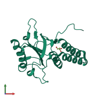
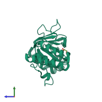
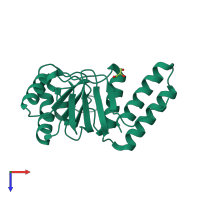
1fy9

X-ray diffraction
2.2Å resolution

CRYSTAL STRUCTURE OF THE HEXA-SUBSTITUTED MUTANT OF THE MOLECULAR CHAPERONIN GROEL APICAL DOMAIN

Released:
Source organism: Escherichia coli
Primary publication:
Stabilization of GroEL minichaperones by core and surface mutations.
J Mol Biol 298 917-26 (2000)
PMID: 10801358

Function and Biology Details

Reaction catalysed:
ATP + H(2)O + a folded polypeptide = ADP + phosphate + an unfolded polypeptide
Biological process:
Cellular component:
  • not assigned

Structure analysis Details

Assembly composition:
monomeric (preferred)
Assembly name:
PDBe Complex ID:
PDB-CPX-141313 (preferred)
Entry contents:
1 distinct polypeptide molecule
Macromolecule:
Chaperonin GroEL Chain: A
Molecule details ›
Chain: A
Length: 193 amino acids
Theoretical weight: 20.85 KDa
Source organism: Escherichia coli
Expression system: Escherichia coli
UniProt:
  • Canonical: P0A6F5 (Residues: 191-376; Coverage: 34%)
Gene names: JW4103, b4143, groEL, groL, mopA
Sequence domains: TCP-1/cpn60 chaperonin family
Structure domains: GroEL

Ligands and Environments

1 bound ligand:
No modified residues

Experiments and Validation Details

Entry percentile scores
X-ray source: LURE BEAMLINE DW32
Spacegroup: P21212
Unit cell:
a: 75.91Å b: 84.52Å c: 35.28Å
α: 90° β: 90° γ: 90°
R-values:
R R work R free
0.265 0.257 0.292
Expression system: Escherichia coli