1g4b

X-ray diffraction
7Å resolution

CRYSTAL STRUCTURES OF THE HSLVU PEPTIDASE-ATPASE COMPLEX REVEAL AN ATP-DEPENDENT PROTEOLYSIS MECHANISM

Released:

Function and Biology Details

Structure analysis Details

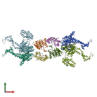
Assembly composition:
hetero 24-mer (preferred)
Assembly name:
PDBe Complex ID:
PDB-CPX-141349 (preferred)
Entry contents:
2 distinct polypeptide molecules
Macromolecules (2 distinct):
ATP-dependent protease ATPase subunit HslU Chains: E, F, K, L
Molecule details ›
Chains: E, F, K, L
Length: 443 amino acids
Theoretical weight: 49.66 KDa
Source organism: Escherichia coli
Expression system: Escherichia coli
UniProt:
  • Canonical: P0A6H5 (Residues: 1-443; Coverage: 100%)
Gene names: JW3902, b3931, hslU, htpI
Sequence domains:
ATP-dependent protease subunit HslV Chains: M, N, O, P
Molecule details ›
Chains: M, N, O, P
Length: 175 amino acids
Theoretical weight: 18.99 KDa
Source organism: Escherichia coli
Expression system: Escherichia coli
UniProt:
  • Canonical: P0A7B8 (Residues: 2-176; Coverage: 99%)
Gene names: JW3903, b3932, hslV, htpO, yiiC
Sequence domains: Proteasome subunit

Ligands and Environments

No bound ligands
No modified residues

Experiments and Validation Details

Entry percentile scores
Spacegroup: P321
Unit cell:
a: 173.376Å b: 173.376Å c: 254.411Å
α: 90° β: 90° γ: 120°
R-values:
R R work R free
0.401 0.401 0.432
Expression system: Escherichia coli