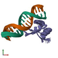
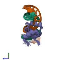
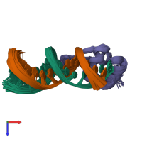
1g4d

Solution NMR

NMR STRUCTURE OF THE MU BACTERIOPHAGE REPRESSOR DNA-BINDING DOMAIN/DNA COMPLEX

Released:
Source organism: Muvirus mu

Function and Biology Details

Biochemical function:
Biological process:
  • not assigned
Cellular component:
  • not assigned

Structure analysis Details

Assembly composition:
hetero trimer (preferred)
PDBe Complex ID:
PDB-CPX-113845 (preferred)
Entry contents:
1 distinct polypeptide molecule
2 distinct DNA molecules
Macromolecules (3 distinct):
Repressor protein c Chain: A
Molecule details ›
Chain: A
Length: 69 amino acids
Theoretical weight: 7.54 KDa
Source organism: Escherichia virus Mu
Expression system: Escherichia coli BL21
UniProt:
  • Canonical: P06019 (Residues: 13-81; Coverage: 35%)
Gene names: Mup01, c, repc
Sequence domains: Mu DNA-binding domain
Structure domains: Winged helix-like DNA-binding domain superfamily/Winged helix DNA-binding domain
5'-D(P*CP*CP*TP*TP*TP*TP*CP*AP*GP*TP*AP*AP*TP*CP*TP*G)-3' Chain: B
Molecule details ›
Chain: B
Length: 16 nucleotides
Theoretical weight: 4.84 KDa
Source organism: Muvirus mu
Expression system: Not provided
5'-D(P*CP*AP*GP*AP*TP*TP*AP*CP*TP*GP*AP*AP*AP*AP*GP*G)-3' Chain: C
Molecule details ›
Chain: C
Length: 16 nucleotides
Theoretical weight: 4.96 KDa
Source organism: Muvirus mu
Expression system: Not provided

Ligands and Environments

No bound ligands
No modified residues

Experiments and Validation Details

Entry percentile scores
Refinement method: simulated annealing
Expression systems:
  • Escherichia coli BL21
  • Not provided