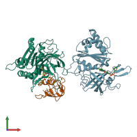
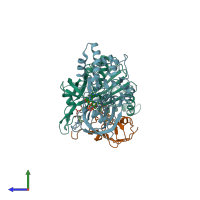
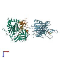
Function and Biology Details
Reaction catalysed:
2 reduced ferredoxin + NADP(+) + H(+) = 2 oxidized ferredoxin + NADPH
Biochemical function:
Biological process:
Cellular component:
- not assigned
Sequence domains:
- Flavoprotein pyridine nucleotide cytochrome reductase
- Ferredoxin-NADP reductase (FNR), nucleotide-binding domain
- Ferredoxin--NADP reductase
- 2Fe-2S ferredoxin-type iron-sulfur binding domain
- Oxidoreductase FAD/NAD(P)-binding
- Flavoprotein pyridine nucleotide cytochrome reductase-like, FAD-binding domain
- FAD-binding domain, ferredoxin reductase-type
- Riboflavin synthase-like beta-barrel
5 more domains
Structure domains:
Structure analysis Details
Assemblies composition:
Assembly name:
PDBe Complex ID:
PDB-CPX-151069 (preferred)
Entry contents:
2 distinct polypeptide molecules
Macromolecules (2 distinct):


