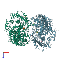
1gqj

X-ray diffraction
1.9Å resolution

Structure of Pseudomonas cellulosa alpha-D-glucuronidase complexed with xylobiose

Released:

Function and Biology Details

Structure analysis Details

Assembly composition:
homo dimer (preferred)
PDBe Complex ID:
PDB-CPX-109885 (preferred)
Entry contents:
1 distinct polypeptide molecule
Macromolecules (2 distinct):
Extracellular xylan exo-alpha-(1->2)-glucuronosidase Chains: A, B
Molecule details ›
Chains: A, B
Length: 708 amino acids
Theoretical weight: 80.44 KDa
Source organism: Cellvibrio japonicus
Expression system: Escherichia coli
UniProt:
  • Canonical: B3PC73 (Residues: 25-732; Coverage: 99%)
Gene names: CJA_2887, gla67A, glcA67A
Sequence domains:
Structure domains:

Ligands and Environments

Carbohydrate polymer : NEW Components: XYP
2 bound ligands:
No modified residues

Experiments and Validation Details

Entry percentile scores
X-ray source: RIGAKU RU200
Spacegroup: P1
Unit cell:
a: 69.561Å b: 74.609Å c: 87.45Å
α: 115.23° β: 93.15° γ: 109.17°
R-values:
R R work R free
0.173 0.172 0.221
Expression system: Escherichia coli