1h9m

X-ray diffraction
1.65Å resolution

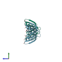
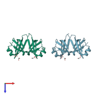
Two crystal structures of the cytoplasmic molybdate-binding protein ModG suggest a novel cooperative binding mechanism and provide insights into ligand-binding specificity. PEG-grown form with molybdate bound

Released:

Function and Biology Details

Structure analysis Details

Assembly composition:
homo trimer (preferred)
PDBe Complex ID:
PDB-CPX-175028 (preferred)
Entry contents:
1 distinct polypeptide molecule
Macromolecule:
Mop domain-containing protein Chains: A, B
Molecule details ›
Chains: A, B
Length: 145 amino acids
Theoretical weight: 14.65 KDa
Source organism: Azotobacter vinelandii
Expression system: Escherichia coli BL21
UniProt:
  • Canonical: Q44529 (Residues: 1-142; Coverage: 100%)
Gene name: modG
Sequence domains: TOBE domain
Structure domains: OB fold (Dihydrolipoamide Acetyltransferase, E2P)

Ligands and Environments

1 bound ligand:
No modified residues

Experiments and Validation Details

Entry percentile scores
X-ray source: ESRF BEAMLINE ID14-2
Spacegroup: R3
Unit cell:
a: 81.96Å b: 81.96Å c: 93.417Å
α: 90° β: 90° γ: 120°
R-values:
R R work R free
0.191 0.191 0.226
Expression system: Escherichia coli BL21