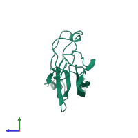
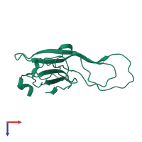
1ifg

X-ray diffraction
2Å resolution

CRYSTAL STRUCTURE OF A MONOMERIC FORM OF GENERAL PROTEASE INHIBITOR, ECOTIN IN ABSENCE OF A PROTEASE

Released:
Source organism: Escherichia coli
Primary publication:
The role of ecotin dimerization in protease inhibition.
J Mol Biol 308 975-91 (2001)
PMID: 11352586

Function and Biology Details

Biological process:
  • not assigned
Cellular component:
  • not assigned

Structure analysis Details

Assembly composition:
monomeric (preferred)
Assembly name:
PDBe Complex ID:
PDB-CPX-150066 (preferred)
Entry contents:
1 distinct polypeptide molecule
Macromolecule:
Ecotin Chain: A
Molecule details ›
Chain: A
Length: 140 amino acids
Theoretical weight: 15.85 KDa
Source organism: Escherichia coli
Expression system: Escherichia coli
UniProt:
  • Canonical: P23827 (Residues: 26-150, 151-162; Coverage: 97%)
Gene names: JW2197, b2209, eco, eti
Sequence domains: Ecotin
Structure domains: Ecotin

Ligands and Environments

No bound ligands
No modified residues

Experiments and Validation Details

Entry percentile scores
X-ray source: SSRL BEAMLINE BL9-1
Spacegroup: P43212
Unit cell:
a: 97.235Å b: 97.235Å c: 37.233Å
α: 90° β: 90° γ: 90°
R-values:
R R work R free
0.248 0.248 0.267
Expression system: Escherichia coli