1imv

X-ray diffraction
2.85Å resolution

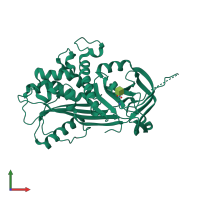
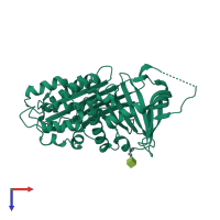
2.85 A crystal structure of PEDF

Released:
Source organism: Homo sapiens
Primary publication:
Crystal structure of human PEDF, a potent anti-angiogenic and neurite growth-promoting factor.
Proc Natl Acad Sci U S A 98 11131-5 (2001)
PMID: 11562499

Function and Biology Details

Structure analysis Details

Assembly composition:
monomeric (preferred)
PDBe Complex ID:
PDB-CPX-153267 (preferred)
Entry contents:
1 distinct polypeptide molecule
Macromolecule:
Pigment epithelium-derived factor Chain: A
Molecule details ›
Chain: A
Length: 398 amino acids
Theoretical weight: 44.33 KDa
Source organism: Homo sapiens
Expression system: Cricetulus griseus
UniProt:
  • Canonical: P36955 (Residues: 21-418; Coverage: 100%)
Gene names: PEDF, PIG35, SERPINF1
Sequence domains: Serpin (serine protease inhibitor)
Structure domains:

Ligands and Environments

1 bound ligand:
No modified residues

Experiments and Validation Details

Entry percentile scores
X-ray source: RIGAKU RU200
Spacegroup: P21212
Unit cell:
a: 176.174Å b: 62.514Å c: 45.406Å
α: 90° β: 90° γ: 90°
R-values:
R R work R free
0.188 0.188 0.227
Expression system: Cricetulus griseus