1j0r

X-ray diffraction
2.5Å resolution

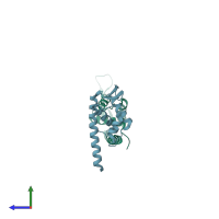
Crystal structure of the replication termination protein mutant C110S

Released:
Source organism: Bacillus subtilis
Primary publication:
The impact of single cysteine residue mutations on the replication terminator protein.
Biochem Biophys Res Commun 310 1096-103 (2003)
PMID: 14559228

Function and Biology Details

Structure analysis Details

Assembly composition:
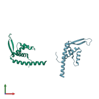
homo dimer (preferred)
PDBe Complex ID:
PDB-CPX-143505 (preferred)
Entry contents:
1 distinct polypeptide molecule
Macromolecule:
Replication termination protein Chains: A, B
Molecule details ›
Chains: A, B
Length: 122 amino acids
Theoretical weight: 14.53 KDa
Source organism: Bacillus subtilis
Expression system: Escherichia coli
UniProt:
  • Canonical: P0CI76 (Residues: 1-122; Coverage: 100%)
Gene names: BSU18490, rtp
Sequence domains: Replication terminator protein
Structure domains: Winged helix-like DNA-binding domain superfamily/Winged helix DNA-binding domain

Ligands and Environments

No bound ligands
No modified residues

Experiments and Validation Details

Entry percentile scores
X-ray source: RIGAKU RU200
Spacegroup: C2
Unit cell:
a: 76.88Å b: 50.94Å c: 69.65Å
α: 90° β: 91.17° γ: 90°
R-values:
R R work R free
0.233 0.233 0.279
Expression system: Escherichia coli