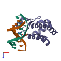
1j3e

X-ray diffraction
2.5Å resolution

Crystal Structure of the E.coli SeqA protein complexed with N6-methyladenine- guanine mismatch DNA

Released:

Function and Biology Details

Structure analysis Details

Assembly composition:
hetero trimer (preferred)
PDBe Complex ID:
PDB-CPX-113984 (preferred)
Entry contents:
1 distinct polypeptide molecule
2 distinct DNA molecules
Macromolecules (3 distinct):
Negative modulator of initiation of replication Chain: A
Molecule details ›
Chain: A
Length: 115 amino acids
Theoretical weight: 12.98 KDa
Source organism: Escherichia coli
Expression system: Escherichia coli
UniProt:
  • Canonical: P0AFY8 (Residues: 71-181; Coverage: 61%)
Gene names: JW0674, b0687, seqA
Sequence domains: SeqA protein C-terminal domain
Structure domains: Replication modulator SeqA, C-terminal DNA-binding domain
5'-D(*AP*AP*GP*GP*(6MA)P*TP*CP*CP*AP*A)-3' Chain: B
Molecule details ›
Chain: B
Length: 10 nucleotides
Theoretical weight: 3.08 KDa
Source organism: Escherichia coli
Expression system: Not provided
5'-D(*TP*TP*GP*GP*AP*GP*CP*CP*TP*T)-3' Chain: C
Molecule details ›
Chain: C
Length: 10 nucleotides
Theoretical weight: 3.05 KDa
Source organism: Escherichia coli
Expression system: Not provided

Ligands and Environments

No bound ligands
1 modified residue:

Experiments and Validation Details

Entry percentile scores
X-ray source: SPRING-8 BEAMLINE BL41XU
Spacegroup: P212121
Unit cell:
a: 33.432Å b: 65.061Å c: 77.176Å
α: 90° β: 90° γ: 90°
R-values:
R R work R free
0.23 0.23 0.296
Expression systems:
  • Escherichia coli
  • Not provided