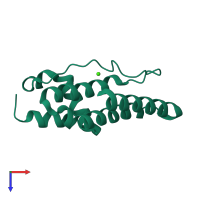
1kp4

X-ray diffraction
1.6Å resolution

CALCIUM-BOUND FORM OF PROKARYOTIC PHOSPHOLIPASE A2

Released:
Source organism: Streptomyces violaceoruber
Primary publication:
The crystal structure of prokaryotic phospholipase A2.
J Biol Chem 277 20059-69 (2002)
PMID: 11897785

Function and Biology Details

Reaction catalysed:
Phosphatidylcholine + H(2)O = 1-acylglycerophosphocholine + a carboxylate
Biochemical function:
Biological process:
Cellular component:
  • not assigned

Structure analysis Details

Assembly composition:
monomeric (preferred)
Assembly name:
PDBe Complex ID:
PDB-CPX-180414 (preferred)
Entry contents:
1 distinct polypeptide molecule
Macromolecule:
Phospholipase A2 Chain: A
Molecule details ›
Chain: A
Length: 122 amino acids
Theoretical weight: 13.57 KDa
Source organism: Streptomyces violaceoruber
Expression system: Streptomyces lividans TK24
UniProt:
  • Canonical: Q6UV28 (Residues: 30-150; Coverage: 96%)
Sequence domains: Prokaryotic phospholipase A2
Structure domains: Phospholipase A2 domain

Ligands and Environments

1 bound ligand:
No modified residues

Experiments and Validation Details

Entry percentile scores
X-ray source: RIGAKU RU300
Spacegroup: P21
Unit cell:
a: 38.28Å b: 54.25Å c: 30.65Å
α: 90° β: 90.2° γ: 90°
R-values:
R R work R free
0.15 0.15 not available
Expression system: Streptomyces lividans TK24