


Function and Biology Details
Biochemical function:
- not assigned
Biological process:
- not assigned
Cellular component:
- not assigned
Structure analysis Details
Assembly composition:
homo dimer (preferred)
Assembly name:
PDBe Complex ID:
PDB-CPX-146149 (preferred)
Entry contents:
1 distinct polypeptide molecule
Macromolecule:
cAMP-dependent protein kinase type II-alpha regulatory subunit
Molecule details ›
Chains: A, B
Length: 46 amino acids
Theoretical weight: 5.4 KDa
Source organism: Mus musculus
Expression system: Escherichia coli
UniProt:
Sequence domains: Regulatory subunit of type II PKA R-subunit
Structure domains: cAMP-dependent protein kinase regulatory subunit, dimerization-anchoring domain
Length: 46 amino acids
Theoretical weight: 5.4 KDa
Source organism: Mus musculus
Expression system: Escherichia coli
UniProt:
- Canonical:
 P12367 (Residues: 1-21, 22-44; Coverage: 11%)
Sequence domains: Regulatory subunit of type II PKA R-subunit
Structure domains: cAMP-dependent protein kinase regulatory subunit, dimerization-anchoring domain
Ligands and Environments
No bound ligands
No modified residues
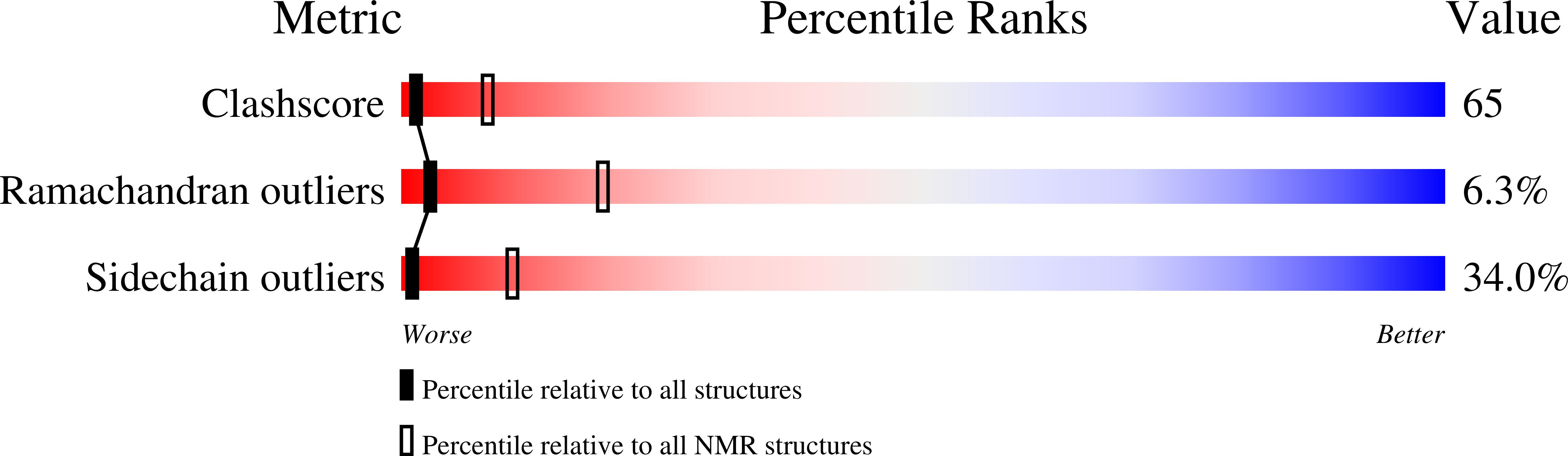
Experiments and Validation Details

Refinement method:
Hybrid distance geometry-dynamical simulated annealing and refinement protocol for monomer structure determination,
with 457 NOE-derived distance restraints (185 intra-residue, i-j=0; 136 sequential, |i-j|=1; 95 medium range, 1<|i-j|<5;
41 long range, |i-j|>4), 19 distance restraints representing hydrogen
bonds (entered as 2 distances each), 25 phi- and 5 chi1-torsion angle restraints.
Molecular dynamical simulated annealing protocol for dimer structure determination, using
505 NOE-derived distance restraints (185 intra-residue, i-j=0; 136 sequential, |i-j|=1;
95 medium range, 1<|i-j|<5; 25 long range, |i-j|>4; 38 inter-molecular; 26 ambiguous),
19 distance restraints representing hydrogen bonds (entered as 2 distances each),
25 phi- and 5 chi1-torsion angle restraints.
Expression system: Escherichia coli