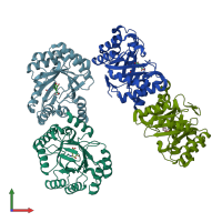
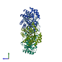
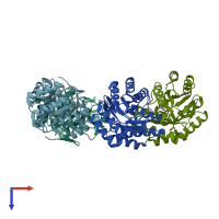
1l9w

X-ray diffraction
2.1Å resolution

CRYSTAL STRUCTURE OF 3-DEHYDROQUINASE FROM SALMONELLA TYPHI COMPLEXED WITH REACTION PRODUCT

Released:

Function and Biology Details

Reaction catalysed:
3-dehydroquinate = 3-dehydroshikimate + H(2)O
Biochemical function:
Cellular component:
  • not assigned

Structure analysis Details

Assembly composition:
homo dimer (preferred)
PDBe Complex ID:
PDB-CPX-150271 (preferred)
Entry contents:
1 distinct polypeptide molecule
Macromolecule:
3-dehydroquinate dehydratase Chains: A, B, C, D
Molecule details ›
Chains: A, B, C, D
Length: 252 amino acids
Theoretical weight: 27.65 KDa
Source organism: Salmonella enterica subsp. enterica serovar Typhi
UniProt:
  • Canonical: P24670 (Residues: 1-252; Coverage: 100%)
Gene names: STY1760, aroD, t1231
Sequence domains: Type I 3-dehydroquinase
Structure domains: Aldolase class I

Ligands and Environments

1 bound ligand:
No modified residues

Experiments and Validation Details

Entry percentile scores
X-ray source: EMBL/DESY, HAMBURG BEAMLINE X31
Spacegroup: P21
Unit cell:
a: 42.61Å b: 158.56Å c: 85.89Å
α: 90° β: 93.61° γ: 90°
R-values:
R R work R free
0.177 0.177 0.245