1ldp

X-ray diffraction
3.1Å resolution

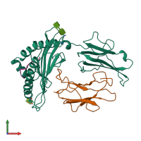
CRYSTAL STRUCTURE OF MURINE MHC CLASS I H-2LD WITH A MIXTURE OF BOUND PEPTIDES

Released:

Function and Biology Details

Structure analysis Details

Assembly composition:
hetero tetramer (preferred)
PDBe Complex ID:
PDB-CPX-134977 (preferred)
Entry contents:
4 distinct polypeptide molecules
Macromolecules (5 distinct):
H-2 class I histocompatibility antigen, L-D alpha chain Chain: H
Molecule details ›
Chain: H
Length: 272 amino acids
Theoretical weight: 31.39 KDa
Source organism: Mus musculus
Expression system: Drosophila melanogaster
UniProt:
  • Canonical: P01897 (Residues: 25-296; Coverage: 81%)
Gene name: H2-L
Sequence domains:
Structure domains:
Beta-2-microglobulin Chain: L
Molecule details ›
Chain: L
Length: 99 amino acids
Theoretical weight: 11.7 KDa
Source organism: Mus musculus
Expression system: Drosophila melanogaster
UniProt:
  • Canonical: P01887 (Residues: 21-119; Coverage: 100%)
Gene name: B2m
Sequence domains: Immunoglobulin C1-set domain
Structure domains: Immunoglobulins
PEPTIDE Chain: P
Molecule details ›
Chain: P
Length: 9 amino acids
Theoretical weight: 744 Da
Source organism: Drosophila melanogaster
PEPTIDE Chain: Q
Molecule details ›
Chain: Q
Length: 9 amino acids
Theoretical weight: 1.06 KDa
Source organism: Drosophila melanogaster

Ligands and Environments

Carbohydrate polymer : NEW Components: NAG, NDG
1 bound ligand:
No modified residues

Experiments and Validation Details

Entry percentile scores
X-ray source: ELLIOTT GX-18
Spacegroup: P2
Unit cell:
a: 72.4Å b: 48.98Å c: 84.06Å
α: 90° β: 97.95° γ: 90°
R-values:
R R work R free
0.221 0.221 0.371
Expression system: Drosophila melanogaster