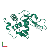
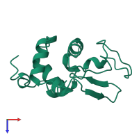
1lmn

X-ray diffraction
1.8Å resolution

THE REFINED CRYSTAL STRUCTURE OF LYSOZYME FROM THE RAINBOW TROUT (ONCORHYNCHUS MYKISS)

Released:
Source organism: Oncorhynchus mykiss
Primary publication:
Refined crystal structure of lysozyme from the rainbow trout (Oncorhynchus mykiss).
Acta Crystallogr D Biol Crystallogr 51 354-67 (1995)
PMID: 15299303

Function and Biology Details

Reaction catalysed:
Hydrolysis of (1->4)-beta-linkages between N-acetylmuramic acid and N-acetyl-D-glucosamine residues in a peptidoglycan and between N-acetyl-D-glucosamine residues in chitodextrins

Structure analysis Details

Assembly composition:
monomeric (preferred)
Assembly name:
PDBe Complex ID:
PDB-CPX-146006 (preferred)
Entry contents:
1 distinct polypeptide molecule
Macromolecule:
Lysozyme C II Chain: A
Molecule details ›
Chain: A
Length: 129 amino acids
Theoretical weight: 14.3 KDa
Source organism: Oncorhynchus mykiss
Expression system: Not provided
UniProt:
  • Canonical: P11941 (Residues: 16-144; Coverage: 100%)
Sequence domains: C-type lysozyme/alpha-lactalbumin family
Structure domains: Lysozyme

Ligands and Environments

No bound ligands
No modified residues

Experiments and Validation Details

Entry percentile scores
Spacegroup: P3121
Unit cell:
a: 76.68Å b: 76.68Å c: 54.46Å
α: 90° β: 90° γ: 120°
R-values:
R R work R free
0.174 not available not available
Expression system: Not provided