Function and Biology Details
Reaction catalysed:
ATP-independent breakage of single-stranded DNA, followed by passage and rejoining
Biochemical function:
Biological process:
Cellular component:
- not assigned
Sequence domains:
- DNA topoisomerase, type IA, central
- TOPRIM domain
- DNA topoisomerase, type IA, active site
- DNA topoisomerase, type IA, central region, subdomain 3
- DNA topoisomerase, type IA, DNA-binding domain
- DNA topoisomerase, type IA, central region, subdomain 2
- DNA topoisomerase, type IA, central region, subdomain 1
- DNA topoisomerase, type IA, domain 2
5 more domains
Structure domain:
Structure analysis Details
Assembly composition:
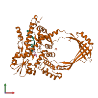
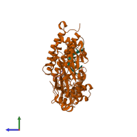
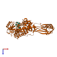
hetero dimer (preferred)
Assembly name:
DNA topoisomerase 1 and DNA (preferred)
PDBe Complex ID:
PDB-CPX-114208 (preferred)
Entry contents:
1 distinct polypeptide molecule
1 distinct DNA molecule
1 distinct DNA molecule
Macromolecules (2 distinct):


