1nfv

X-ray diffraction
1.95Å resolution

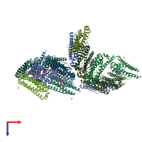
X-ray structure of Desulfovibrio desulfuricans bacterioferritin: the diiron centre in different catalytic states (as-isolated structure)

Released:

Function and Biology Details

Reaction catalysed:
4 Fe(2+) + 4 H(+) + O(2) = 4 Fe(3+) + 2 H(2)O
Biochemical function:
Cellular component:

Structure analysis Details

Assemblies composition:
homo hexadecamer (preferred)
homo 24-mer
Assembly name:
PDBe Complex ID:
PDB-CPX-188112 (preferred)
Entry contents:
1 distinct polypeptide molecule
Macromolecule:
Bacterioferritin Chains: A, B, C, D, E, F, G, H, I, J, K, L, M, N, O, P
Molecule details ›
Chains: A, B, C, D, E, F, G, H, I, J, K, L, M, N, O, P
Length: 179 amino acids
Theoretical weight: 19.91 KDa
Source organism: Desulfovibrio desulfuricans
UniProt:
  • Canonical: Q93PP9 (Residues: 1-179; Coverage: 100%)
Gene names: Ddes_1387, bfr
Sequence domains: Ferritin-like domain
Structure domains: Ferritin

Ligands and Environments

No modified residues

Experiments and Validation Details

Entry percentile scores
X-ray source: ESRF BEAMLINE BM14, ESRF BEAMLINE ID14-2
Spacegroup: P213
Unit cell:
a: 225.3Å b: 225.3Å c: 225.3Å
α: 90° β: 90° γ: 90°
R-values:
R R work R free
0.214 0.214 0.254