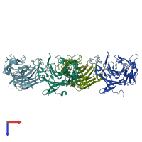
1o4z

X-ray diffraction
2.3Å resolution

THE THREE-DIMENSIONAL STRUCTURE OF BETA-AGARASE B FROM ZOBELLIA GALACTANIVORANS

Released:
Source organism: Zobellia galactanivorans
Primary publication:
The three-dimensional structures of two beta-agarases.
J Biol Chem 278 47171-80 (2003)
PMID: 12970344

Function and Biology Details

Reaction catalysed:
Hydrolysis of (1->4)-beta-D-galactosidic linkages in agarose, giving the tetramer as the predominant product
Biochemical function:
Biological process:
Cellular component:

Structure analysis Details

Assembly composition:
homo dimer (preferred)
Assembly name:
PDBe Complex ID:
PDB-CPX-193171 (preferred)
Entry contents:
1 distinct polypeptide molecule
Macromolecule:
Beta-agarase B Chains: A, B, C, D
Molecule details ›
Chains: A, B, C, D
Length: 346 amino acids
Theoretical weight: 40.06 KDa
Source organism: Zobellia galactanivorans
Expression system: Escherichia coli
UniProt:
  • Canonical: Q9RGX8 (Residues: 19-353; Coverage: 100%)
Gene names: agaB, zobellia_3573
Structure domains: Jelly Rolls

Ligands and Environments

3 bound ligands:
No modified residues

Experiments and Validation Details

Entry percentile scores
X-ray source: ESRF BEAMLINE ID14-2
Spacegroup: P21
Unit cell:
a: 72.448Å b: 105.35Å c: 97.369Å
α: 90° β: 94.13° γ: 90°
R-values:
R R work R free
0.169 0.166 0.224
Expression system: Escherichia coli