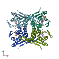
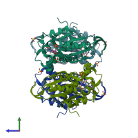
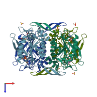
1o5o

X-ray diffraction
2.3Å resolution

Crystal structure of Uracil phosphoribosyltransferase (TM0721) from Thermotoga maritima at 2.30 A resolution

Released:
Source organism: Thermotoga maritima
Entry author: Joint Center for Structural Genomics (JCSG)

Function and Biology Details

Reaction catalysed:
UMP + diphosphate = uracil + 5-phospho-alpha-D-ribose 1-diphosphate
Biochemical function:
Biological process:
Cellular component:

Structure analysis Details

Assembly composition:
homo tetramer (preferred)
Entry contents:
1 distinct polypeptide molecule
Macromolecule:
Uracil phosphoribosyltransferase Chains: A, B, C, D
Molecule details ›
Chains: A, B, C, D
Length: 221 amino acids
Theoretical weight: 24.81 KDa
Source organism: Thermotoga maritima
Expression system: Escherichia coli
UniProt:
  • Canonical: Q9WZI0 (Residues: 1-209; Coverage: 100%)
Gene names: TM_0721, upp
Sequence domains: Uracil phosphoribosyltransferase
Structure domains: Rossmann fold

Ligands and Environments

2 bound ligands:
No modified residues

Experiments and Validation Details

Entry percentile scores
X-ray source: APS BEAMLINE 14-BM-C
Spacegroup: C2
Unit cell:
a: 134.078Å b: 87.406Å c: 90.826Å
α: 90° β: 115.29° γ: 90°
R-values:
R R work R free
0.17 0.167 0.236
Expression system: Escherichia coli