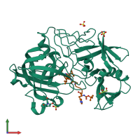
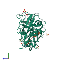
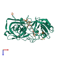
1oex

X-ray diffraction
1.1Å resolution

Atomic Resolution Structure of Endothiapepsin in Complex with a Hydroxyethylene Transition State Analogue Inhibitor H261

Released:

Function and Biology Details

Reaction catalysed:
Hydrolysis of proteins with specificity similar to that of pepsin A, prefers hydrophobic residues at P1 and P1', but does not cleave 14-Ala-|-Leu-15 in the B chain of insulin or Z-Glu-Tyr. Clots milk.
Biological process:
Cellular component:
  • not assigned

Structure analysis Details

Assembly composition:
hetero dimer (preferred)
PDBe Complex ID:
PDB-CPX-145951 (preferred)
Entry contents:
2 distinct polypeptide molecules
Macromolecules (2 distinct):
Endothiapepsin Chain: A
Molecule details ›
Chain: A
Length: 329 amino acids
Theoretical weight: 33.8 KDa
Source organism: Cryphonectria parasitica
UniProt:
  • Canonical: P11838 (Residues: 90-419; Coverage: 83%)
Gene names: EAPA, EPN-1
Sequence domains: Eukaryotic aspartyl protease
Structure domains: Acid Proteases
INHIBITOR H261 Chain: B
Molecule details ›
Chain: B
Length: 8 amino acids
Theoretical weight: 1.04 KDa
Source organism: synthetic construct
Expression system: Not provided

Ligands and Environments

2 bound ligands:
1 modified residue:

Experiments and Validation Details

Entry percentile scores
X-ray source: EMBL/DESY, HAMBURG BEAMLINE BW7B
Spacegroup: P21
Unit cell:
a: 42.58Å b: 74.51Å c: 42.35Å
α: 90° β: 97.56° γ: 90°
R-values:
R R work R free
0.14 not available 0.178
Expression system: Not provided