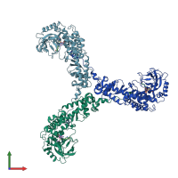
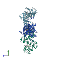
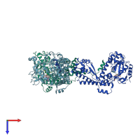
1ozv

X-ray diffraction
2.65Å resolution

Crystal structure of the SET domain of LSMT bound to Lysine and AdoHcy

Released:
Source organism: Pisum sativum

Function and Biology Details

Reactions catalysed:
3 S-adenosyl-L-methionine + [ribulose-bisphosphate carboxylase]-L-lysine = 3 S-adenosyl-L-homocysteine + [ribulose-bisphosphate carboxylase]-N(6),N(6),N(6)-trimethyl-L-lysine
3 S-adenosyl-L-methionine + [fructose-bisphosphate aldolase]-L-lysine = 3 S-adenosyl-L-homocysteine + [fructose-bisphosphate aldolase]-N(6),N(6),N(6)-trimethyl-L-lysine

Structure analysis Details

Assemblies composition:
monomeric (preferred)
homo trimer
PDBe Complex ID:
PDB-CPX-174981 (preferred)
Entry contents:
1 distinct polypeptide molecule
Macromolecule:
Ribulose-1,5 bisphosphate carboxylase/oxygenase large subunit N-methyltransferase, chloroplastic Chains: A, B, C
Molecule details ›
Chains: A, B, C
Length: 444 amino acids
Theoretical weight: 50.63 KDa
Source organism: Pisum sativum
Expression system: Escherichia coli BL21
UniProt:
  • Canonical: Q43088 (Residues: 46-482; Coverage: 89%)
Gene name: RBCMT
Sequence domains: Rubisco LSMT substrate-binding
Structure domains:

Ligands and Environments


Cofactor: Ligand SAH 3 x SAH
1 bound ligand:
No modified residues

Experiments and Validation Details

Entry percentile scores
X-ray source: RIGAKU RU200
Spacegroup: I222
Unit cell:
a: 131.96Å b: 156.9Å c: 267.55Å
α: 90° β: 90° γ: 90°
R-values:
R R work R free
0.229 0.227 0.266
Expression system: Escherichia coli BL21