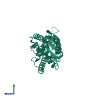
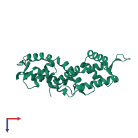
1p4x

X-ray diffraction
2.2Å resolution

Crystal structure of SarS protein from Staphylococcus Aureus

Released:
Source organism: Staphylococcus aureus
Primary publication:
Crystal structure of the SarS protein from Staphylococcus aureus.
J Bacteriol 185 4219-25 (2003)
PMID: 12837797

Function and Biology Details

Structure analysis Details

Assembly composition:
monomeric (preferred)
PDBe Complex ID:
PDB-CPX-173530 (preferred)
Entry contents:
1 distinct polypeptide molecule
Macromolecule:
HTH-type transcriptional regulator SarS Chain: A
Molecule details ›
Chain: A
Length: 250 amino acids
Theoretical weight: 29.94 KDa
Source organism: Staphylococcus aureus
Expression system: Escherichia coli
UniProt:
  • Canonical: Q2G1N7 (Residues: 1-250; Coverage: 100%)
Gene names: SAOUHSC_00070, sarH1, sarS
Sequence domains: MarR family
Structure domains: Winged helix-like DNA-binding domain superfamily/Winged helix DNA-binding domain

Ligands and Environments

No bound ligands
No modified residues

Experiments and Validation Details

Entry percentile scores
X-ray source: RIGAKU
Spacegroup: P6522
Unit cell:
a: 84.887Å b: 84.887Å c: 195.743Å
α: 90° β: 90° γ: 120°
R-values:
R R work R free
0.24 0.24 0.285
Expression system: Escherichia coli