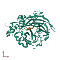
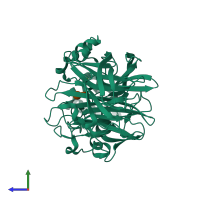
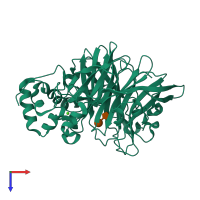
1pt2

X-ray diffraction
2.1Å resolution

Crystal structure of levansucrase (E342A) complexed with sucrose

Released:
Source organism: Bacillus subtilis
Primary publication:
Structural framework of fructosyl transfer in Bacillus subtilis levansucrase.
Nat Struct Biol 10 935-41 (2003)
PMID: 14517548

Function and Biology Details

Reaction catalysed:
Sucrose + (6)-beta-D-fructofuranosyl-(2->)(n) alpha-D-glucopyranoside = glucose + (6)-beta-D-fructofuranosyl-(2->)(n+1) alpha-D-glucopyranoside
Biochemical function:
Biological process:
Cellular component:

Structure analysis Details

Assembly composition:
monomeric (preferred)
Assembly name:
PDBe Complex ID:
PDB-CPX-138729 (preferred)
Entry contents:
1 distinct polypeptide molecule
Macromolecules (2 distinct):
Levansucrase Chain: A
Molecule details ›
Chain: A
Length: 447 amino acids
Theoretical weight: 50.26 KDa
Source organism: Bacillus subtilis
Expression system: Escherichia coli BL21(DE3)
UniProt:
  • Canonical: P05655 (Residues: 30-473; Coverage: 100%)
Gene names: BSU34450, sacB
Sequence domains: Levansucrase/Invertase
Structure domains: Glycosyl hydrolase domain; family 43

Ligands and Environments

Carbohydrate polymer : NEW Components: GLC, FRU
1 bound ligand:
No modified residues

Experiments and Validation Details

Entry percentile scores
X-ray source: ENRAF-NONIUS FR591
Spacegroup: P212121
Unit cell:
a: 51.241Å b: 66.951Å c: 123.956Å
α: 90° β: 90° γ: 90°
R-values:
R R work R free
0.191 0.189 0.232
Expression system: Escherichia coli BL21(DE3)