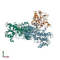
Function and Biology Details
Reaction catalysed:
NADPH + NAD(+) + H(+)(Side 1) = NADP(+) + H(+)(Side 2) + NADH
Biochemical function:
Biological process:
Cellular component:
Sequence domains:
- Alanine dehydrogenase/pyridine nucleotide transhydrogenase, NAD(H)-binding domain
- Alanine dehydrogenase/pyridine nucleotide transhydrogenase, N-terminal
- Alanine dehydrogenase/pyridine nucleotide transhydrogenase, conserved site-2
- NAD(P)-binding domain superfamily
- Alanine dehydrogenase/NAD(P) transhydrogenase, conserved site-1
- NADP transhydrogenase beta-like domain
- DHS-like NAD/FAD-binding domain superfamily
Structure analysis Details
Assembly composition:
hetero trimer (preferred)
Assembly name:
NAD(P) transhydrogenase (preferred)
PDBe Complex ID:
PDB-CPX-173910 (preferred)
Entry contents:
2 distinct polypeptide molecules
Macromolecules (2 distinct):


