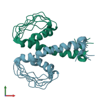
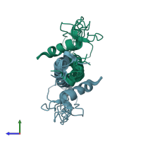
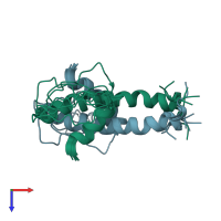
Function and Biology Details
Reaction catalysed:
Propanoyl-CoA + 6 (2S)-methylmalonyl-CoA + 6 NADPH = 6-deoxyerythronolide B + 7 CoA + 6 CO(2) + H(2)O + 6 NADP(+)
Biochemical function:
Biological process:
- not assigned
Cellular component:
- not assigned
Sequence domains:
Structure domain:
Structure analysis Details
Assembly composition:
homo dimer (preferred)
Assembly name:
PDBe Complex ID:
PDB-CPX-169840 (preferred)
Entry contents:
1 distinct polypeptide molecule
Macromolecule:
Ligands and Environments
No bound ligands
No modified residues
Experiments and Validation Details
Refinement method:
simulated annealing
Expression system: Escherichia coli


