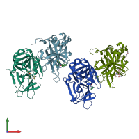
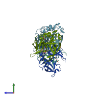
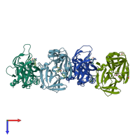
1qa7

X-ray diffraction
1.9Å resolution

CRYSTAL COMPLEX OF THE 3C PROTEINASE FROM HEPATITIS A VIRUS WITH ITS INHIBITOR AND IMPLICATIONS FOR THE POLYPROTEIN PROCESSING IN HAV

Released:

Function and Biology Details

Reactions catalysed:
Nucleoside triphosphate + RNA(n) = diphosphate + RNA(n+1)
Selective cleavage of Gln-|-Gly bond in the poliovirus polyprotein. In other picornavirus reactions Glu may be substituted for Gln, and Ser or Thr for Gly.
NTP + H(2)O = NDP + phosphate
Biological process:
Cellular component:
  • not assigned

Structure analysis Details

Assemblies composition:
monomeric (preferred)
homo tetramer
homo dimer
Assembly name:
PDBe Complex ID:
PDB-CPX-140225 (preferred)
Entry contents:
1 distinct polypeptide molecule
Macromolecule:
Protease 3C Chains: A, B, C, D
Molecule details ›
Chains: A, B, C, D
Length: 217 amino acids
Theoretical weight: 23.83 KDa
Source organism: Hepatovirus A
Expression system: Escherichia coli
UniProt:
  • Canonical: P08617 (Residues: 1520-1736; Coverage: 10%)
Sequence domains: 3C cysteine protease (picornain 3C)
Structure domains: Trypsin-like serine proteases

Ligands and Environments

3 bound ligands:
No modified residues

Experiments and Validation Details

Entry percentile scores
X-ray source: SSRL BEAMLINE BL7-1
Spacegroup: P21
Unit cell:
a: 50.562Å b: 78.357Å c: 105.287Å
α: 90° β: 97.5° γ: 90°
R-values:
R R work R free
0.214 0.204 0.298
Expression system: Escherichia coli