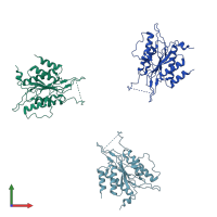
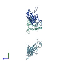
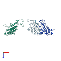
1qc9

X-ray diffraction
3Å resolution

THE CRYSTALLOGRAPHIC STRUCTURE OF RESTRICTION ENDONUCLEASE ECO RI AT 3.3 A IN THE ABSENSE OF DNA

Released:
Source organism: Escherichia coli
Entry authors: Chandrasekhar K, Horvath MM, Samudzi C, Choi J, Rosenberg JM

Function and Biology Details

Reaction catalysed:
Endonucleolytic cleavage of DNA to give specific double-stranded fragments with terminal 5'-phosphates
Biochemical function:
Cellular component:
  • not assigned

Structure analysis Details

Assembly composition:
homo dimer (preferred)
PDBe Complex ID:
PDB-CPX-132789 (preferred)
Entry contents:
1 distinct polypeptide molecule
Macromolecule:
Type II restriction enzyme EcoRI Chains: A, B, C
Molecule details ›
Chains: A, B, C
Length: 276 amino acids
Theoretical weight: 30.97 KDa
Source organism: Escherichia coli
UniProt:
  • Canonical: P00642 (Residues: 2-277; Coverage: 100%)
Gene name: ecoRIR
Sequence domains: Restriction endonuclease EcoRI
Structure domains: Eco RI Endonuclease, subunit A

Ligands and Environments

No bound ligands
No modified residues

Experiments and Validation Details

Entry percentile scores
X-ray source: RIGAKU RU200
Spacegroup: C2
Unit cell:
a: 208.68Å b: 127.35Å c: 49.87Å
α: 90° β: 98.57° γ: 90°
R-values:
R R work R free
0.251 0.251 0.295