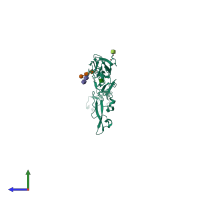
1qub

X-ray diffraction
2.7Å resolution

CRYSTAL STRUCTURE OF THE GLYCOSYLATED FIVE-DOMAIN HUMAN BETA2-GLYCOPROTEIN I PURIFIED FROM BLOOD PLASMA

Released:

Function and Biology Details

Structure analysis Details

Assembly composition:
monomeric (preferred)
Assembly name:
PDBe Complex ID:
PDB-CPX-136130 (preferred)
Entry contents:
1 distinct polypeptide molecule
Macromolecules (3 distinct):
Beta-2-glycoprotein 1 Chain: A
Molecule details ›
Chain: A
Length: 319 amino acids
Theoretical weight: 35.48 KDa
Source organism: Homo sapiens
UniProt:
  • Canonical: P02749 (Residues: 20-345; Coverage: 98%)
Gene names: APOH, B2G1
Sequence domains:
Structure domains: Complement Module, domain 1

Ligands and Environments

Carbohydrate polymer : NEW Components: NAG, MAN
Carbohydrate polymer : NEW Components: NAG
1 bound ligand:
No modified residues

Experiments and Validation Details

Entry percentile scores
X-ray source: ESRF BEAMLINE ID14-4
Spacegroup: C2221
Unit cell:
a: 161.17Å b: 166.49Å c: 114.51Å
α: 90° β: 90° γ: 90°
R-values:
R R work R free
0.25 0.249 0.269