


Function and Biology Details
Reaction catalysed:
Endohydrolysis of the N-glycosidic bond at one specific adenosine on the28S rRNA.
Biochemical function:
Biological process:
Cellular component:
Sequence domains:
Structure domains:
Structure analysis Details
Assemblies composition:
Assembly name:
Shiga toxin (preferred)
PDBe Complex ID:
PDB-CPX-181371 (preferred)
Entry contents:
2 distinct polypeptide molecules
Macromolecules (2 distinct):
Shiga toxin subunit A
Molecule details ›
Chains: A, L
Length: 293 amino acids
Theoretical weight: 32.26 KDa
Source organism: Shigella dysenteriae
Expression system: Escherichia coli
UniProt:
Sequence domains: Ribosome inactivating protein
Structure domains:
Length: 293 amino acids
Theoretical weight: 32.26 KDa
Source organism: Shigella dysenteriae
Expression system: Escherichia coli
UniProt:
- Canonical:
 Q9FBI2 (Residues: 23-315; Coverage: 100%)
Sequence domains: Ribosome inactivating protein
Structure domains:
Shiga toxin subunit B
Molecule details ›
Chains: B, C, D, E, F, G, H, I, J, K
Length: 69 amino acids
Theoretical weight: 7.7 KDa
Source organism: Shigella dysenteriae
Expression system: Escherichia coli
UniProt:
Sequence domains: Shiga-like toxin beta subunit
Structure domains: OB fold (Dihydrolipoamide Acetyltransferase, E2P)
Length: 69 amino acids
Theoretical weight: 7.7 KDa
Source organism: Shigella dysenteriae
Expression system: Escherichia coli
UniProt:
- Canonical: Q7BQ98 (Residues: 21-89; Coverage: 100%)
Sequence domains: Shiga-like toxin beta subunit
Structure domains: OB fold (Dihydrolipoamide Acetyltransferase, E2P)
Ligands and Environments
No bound ligands
No modified residues
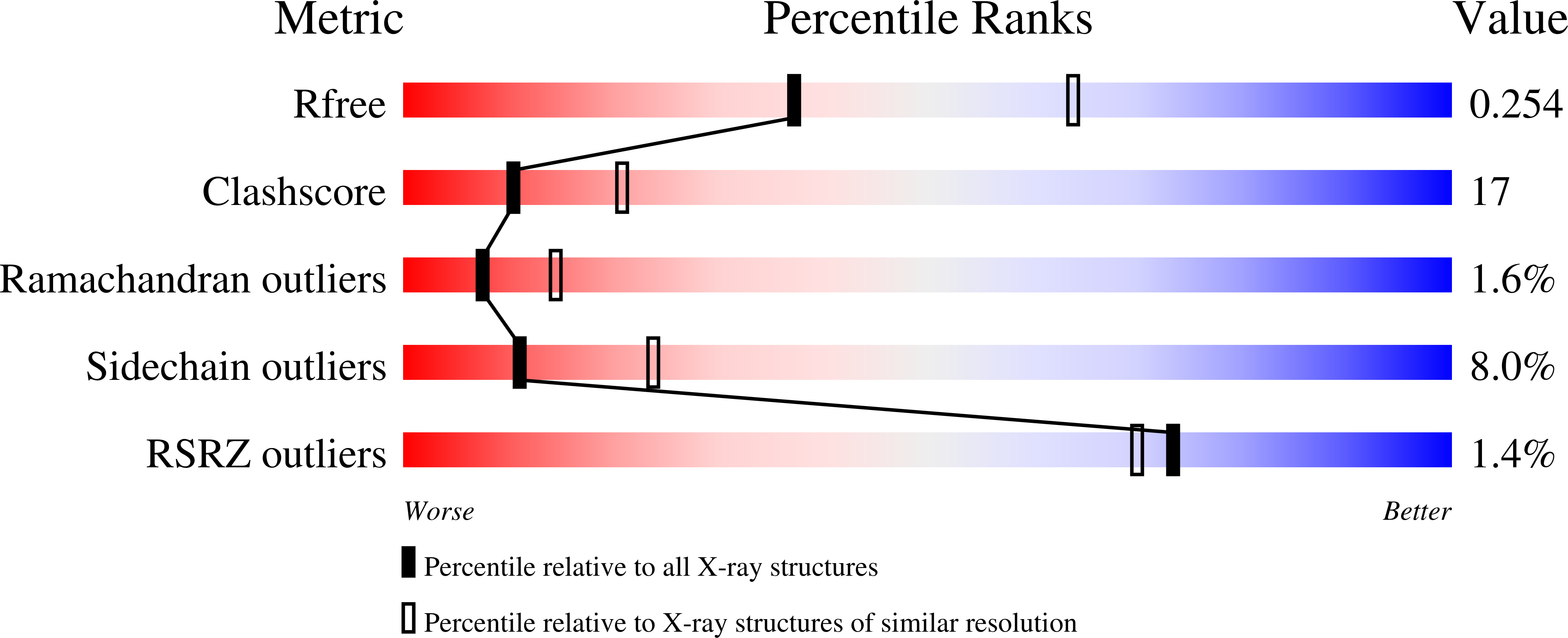
Experiments and Validation Details
wwPDB Validation report is not available for this entry.
X-ray source:
PHOTON FACTORY BEAMLINE BL-6A
Spacegroup:
P212121
Expression system: Escherichia coli
