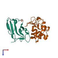
1rjc

X-ray diffraction
1.4Å resolution

Crystal structure of the camelid single domain antibody cAb-Lys2 in complex with hen egg white lysozyme

Released:

Function and Biology Details

Reaction catalysed:
Hydrolysis of (1->4)-beta-linkages between N-acetylmuramic acid and N-acetyl-D-glucosamine residues in a peptidoglycan and between N-acetyl-D-glucosamine residues in chitodextrins

Structure analysis Details

Assembly composition:
hetero dimer (preferred)
PDBe Complex ID:
PDB-CPX-208247 (preferred)
Entry contents:
2 distinct polypeptide molecules
Macromolecules (2 distinct):
camelid heavy chain antibody Chain: A
Molecule details ›
Chain: A
Length: 137 amino acids
Theoretical weight: 14.9 KDa
Source organism: Camelus dromedarius
Expression system: Escherichia coli
Structure domains: Immunoglobulins
Lysozyme C Chain: B
Molecule details ›
Chain: B
Length: 129 amino acids
Theoretical weight: 14.33 KDa
Source organism: Gallus gallus
UniProt:
  • Canonical: P00698 (Residues: 19-147; Coverage: 100%)
Gene name: LYZ
Sequence domains: C-type lysozyme/alpha-lactalbumin family
Structure domains: Lysozyme

Ligands and Environments

2 bound ligands:
No modified residues

Experiments and Validation Details

Entry percentile scores
X-ray source: EMBL/DESY, HAMBURG BEAMLINE BW7B
Spacegroup: P212121
Unit cell:
a: 50.02Å b: 68.36Å c: 72.05Å
α: 90° β: 90° γ: 90°
R-values:
R R work R free
0.22 0.22 0.235
Expression system: Escherichia coli