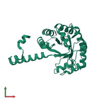
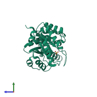
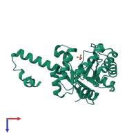
1s2w

X-ray diffraction
1.69Å resolution

Crystal structure of phosphoenolpyruvate mutase in high ionic strength

Released:
Source organism: Mytilus edulis
Primary publication:
Conformational flexibility of PEP mutase.
Biochemistry 43 4447-53 (2004)
PMID: 15078090

Function and Biology Details

Structure analysis Details

Assemblies composition:
monomeric (preferred)
homo dimer
PDBe Complex ID:
PDB-CPX-157566 (preferred)
Entry contents:
1 distinct polypeptide molecule
Macromolecule:
Phosphoenolpyruvate phosphomutase Chain: A
Molecule details ›
Chain: A
Length: 295 amino acids
Theoretical weight: 32.95 KDa
Source organism: Mytilus edulis
Expression system: Escherichia coli BL21(DE3)
UniProt:
  • Canonical: P56839 (Residues: 1-295; Coverage: 100%)
Sequence domains: Phosphoenolpyruvate phosphomutase
Structure domains: Phosphoenolpyruvate-binding domains

Ligands and Environments

1 bound ligand:
No modified residues

Experiments and Validation Details

Entry percentile scores
X-ray source: APS BEAMLINE 17-BM
Spacegroup: C2221
Unit cell:
a: 76.632Å b: 116.556Å c: 72.818Å
α: 90° β: 90° γ: 90°
R-values:
R R work R free
0.183 0.178 0.207
Expression system: Escherichia coli BL21(DE3)