Function and Biology Details
Reaction catalysed:
Catalyzes the rearrangement of -S-S- bonds in proteins
Biochemical function:
Biological process:
Cellular component:
Sequence domains:
Structure domains:
Structure analysis Details
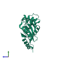
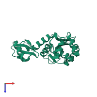
Assembly composition:
homo dimer (preferred)
Assembly name:
Thiol:disulfide interchange protein DsbC (preferred)
PDBe Complex ID:
PDB-CPX-142455 (preferred)
Entry contents:
1 distinct polypeptide molecule
Macromolecule:
Ligands and Environments
No bound ligands
No modified residues
Experiments and Validation Details
X-ray source:
EMBL/DESY, HAMBURG BEAMLINE X11
Spacegroup:
C2221
Expression system: Escherichia coli


