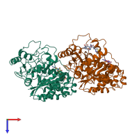
1tvk

Electron crystallography
2.89Å resolution

The binding mode of epothilone A on a,b-tubulin by electron crystallography

Released:

Function and Biology Details

Structure analysis Details

Assembly composition:
hetero dimer (preferred)
PDBe Complex ID:
PDB-CPX-173621 (preferred)
Entry contents:
2 distinct polypeptide molecules
Macromolecules (2 distinct):
Tubulin alpha-1D chain Chain: A
Molecule details ›
Chain: A
Length: 440 amino acids
Theoretical weight: 48.87 KDa
Source organism: Bos taurus
UniProt:
  • Canonical: Q2HJ86 (Residues: 1-440; Coverage: 97%)
Gene name: TUBA1D
Sequence domains:
Structure domains:
Tubulin beta-2B chain Chain: B
Molecule details ›
Chain: B
Length: 427 amino acids
Theoretical weight: 47.94 KDa
Source organism: Bos taurus
UniProt:
  • Canonical: Q6B856 (Residues: 1-427; Coverage: 96%)
Gene name: TUBB2B
Sequence domains:
Structure domains:

Ligands and Environments

3 bound ligands:
No modified residues

Experiments and Validation Details

Entry percentile scores
Resolution: 2.89Å