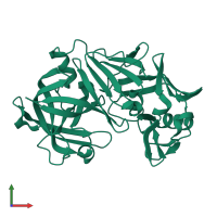
Function and Biology Details
Reaction catalysed:
Hydrolysis of proteins with broad specificity similar to that of pepsin A, preferring hydrophobic residues at P1 and P1'. Clots milk and activates trypsinogen. Does not cleave 4-Gln-|-His-5, but does cleave 10-His-|-Leu-11 and 12-Val-|-Glu-13 in B chain of insulin.
Biochemical function:
Biological process:
Cellular component:
- not assigned
Structure analysis Details
Assembly composition:
monomeric (preferred)
Assembly name:
Rhizopuspepsin (preferred)
PDBe Complex ID:
PDB-CPX-138855 (preferred)
Entry contents:
1 distinct polypeptide molecule
Macromolecule:
Ligands and Environments
No bound ligands
No modified residues
Experiments and Validation Details
X-ray source:
RIGAKU RU200
Spacegroup:
P212121


