1vq2

X-ray diffraction
2.2Å resolution

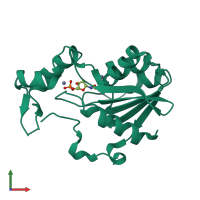
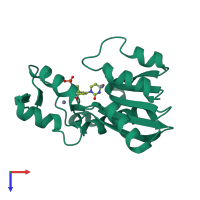
CRYSTAL STRUCTURE OF T4-BACTERIOPHAGE DEOXYCYTIDYLATE DEAMINASE, MUTANT R115E

Released:

Function and Biology Details

Structure analysis Details

Assemblies composition:
homo dimer (preferred)
homo hexamer
PDBe Complex ID:
PDB-CPX-147577 (preferred)
Entry contents:
1 distinct polypeptide molecule
Macromolecule:
Deoxycytidylate deaminase Chain: A
Molecule details ›
Chain: A
Length: 193 amino acids
Theoretical weight: 21.2 KDa
Source organism: Escherichia virus T4
Expression system: Escherichia coli
UniProt:
  • Canonical: P16006 (Residues: 1-193; Coverage: 100%)
Gene name: CD
Sequence domains: Cytidine and deoxycytidylate deaminase zinc-binding region
Structure domains: Cytidine Deaminase, domain 2

Ligands and Environments

2 bound ligands:
No modified residues

Experiments and Validation Details

Entry percentile scores
X-ray source: NSLS BEAMLINE X12C
Spacegroup: P6322
Unit cell:
a: 114.528Å b: 114.528Å c: 76.95Å
α: 90° β: 90° γ: 120°
R-values:
R R work R free
0.218 0.218 0.245
Expression system: Escherichia coli