

-1,4-DIMERCAPTOBUTANE-2,3-DIOL</span>;</li> <li class='image_legend_li'>1 copy of <span class='highlight'>3-{2,6,8-TRIOXO-9-[(2R,3S,4R)-2,3,4,5-TETRAHYDROXYPENTYL]-1,2,3,6,8,9-HEXAHYDRO-7H-PURIN-7-YL}PROPYL DIHYDROGEN PHOSPHATE</span>;</li> <li class='image_legend_li'>5 copies of <span class='highlight'>POTASSIUM ION</span>;</li> <li class='image_legend_li'>1 copy of <span class='highlight'>3-{2,6,8-TRIOXO-9-[(2S,3R,4R)-2,3,4,5-TETRAHYDROXYPENTYL]-1,2,3,6,8,9-HEXAHYDRO-7H-PURIN-7-YL}PROPYL DIHYDROGEN PHOSPHATE</span>;</li> <li class='image_legend_li'>3 copies of <span class='highlight'>(2S,3S)-1,4-DIMERCAPTOBUTANE-2,3-DIOL</span>;</li> <li class='image_legend_li'>1 copy of <span class='highlight'>3-{2,6,8-TRIOXO-9-[(2S,3S,4R)-2,3,4,5-TETRAHYDROXYPENTYL]-1,2,3,6,8,9-HEXAHYDRO-7H-PURIN-7-YL}PROPYL DIHYDROGEN PHOSPHATE</span>;</li> <li class='image_legend_li'>2 copies of <span class='highlight'>3-{2,6,8-TRIOXO-9-[(2R,3R,4R)-2,3,4,5-TETRAHYDROXYPENTYL]-1,2,3,6,8,9-HEXAHYDRO-7H-PURIN-7-YL}PROPYL DIHYDROGEN PHOSPHATE</span>;</li> <li class='image_legend_li'>1 copy of <span class='highlight'>(4S,5S)-1,2-DITHIANE-4,5-DIOL</span>;</li> <li class='image_legend_li'>1 copy of <span class='highlight'>water</span>.</li></ul>")
-1,4-DIMERCAPTOBUTANE-2,3-DIOL</span>;</li> <li class='image_legend_li'>1 copy of <span class='highlight'>3-{2,6,8-TRIOXO-9-[(2R,3S,4R)-2,3,4,5-TETRAHYDROXYPENTYL]-1,2,3,6,8,9-HEXAHYDRO-7H-PURIN-7-YL}PROPYL DIHYDROGEN PHOSPHATE</span>;</li> <li class='image_legend_li'>5 copies of <span class='highlight'>POTASSIUM ION</span>;</li> <li class='image_legend_li'>1 copy of <span class='highlight'>3-{2,6,8-TRIOXO-9-[(2S,3R,4R)-2,3,4,5-TETRAHYDROXYPENTYL]-1,2,3,6,8,9-HEXAHYDRO-7H-PURIN-7-YL}PROPYL DIHYDROGEN PHOSPHATE</span>;</li> <li class='image_legend_li'>3 copies of <span class='highlight'>(2S,3S)-1,4-DIMERCAPTOBUTANE-2,3-DIOL</span>;</li> <li class='image_legend_li'>1 copy of <span class='highlight'>3-{2,6,8-TRIOXO-9-[(2S,3S,4R)-2,3,4,5-TETRAHYDROXYPENTYL]-1,2,3,6,8,9-HEXAHYDRO-7H-PURIN-7-YL}PROPYL DIHYDROGEN PHOSPHATE</span>;</li> <li class='image_legend_li'>2 copies of <span class='highlight'>3-{2,6,8-TRIOXO-9-[(2R,3R,4R)-2,3,4,5-TETRAHYDROXYPENTYL]-1,2,3,6,8,9-HEXAHYDRO-7H-PURIN-7-YL}PROPYL DIHYDROGEN PHOSPHATE</span>;</li> <li class='image_legend_li'>1 copy of <span class='highlight'>(4S,5S)-1,2-DITHIANE-4,5-DIOL</span>;</li> <li class='image_legend_li'>1 copy of <span class='highlight'>water</span>.</li></ul>")
-1,4-DIMERCAPTOBUTANE-2,3-DIOL</span>;</li> <li class='image_legend_li'>1 copy of <span class='highlight'>3-{2,6,8-TRIOXO-9-[(2R,3S,4R)-2,3,4,5-TETRAHYDROXYPENTYL]-1,2,3,6,8,9-HEXAHYDRO-7H-PURIN-7-YL}PROPYL DIHYDROGEN PHOSPHATE</span>;</li> <li class='image_legend_li'>5 copies of <span class='highlight'>POTASSIUM ION</span>;</li> <li class='image_legend_li'>1 copy of <span class='highlight'>3-{2,6,8-TRIOXO-9-[(2S,3R,4R)-2,3,4,5-TETRAHYDROXYPENTYL]-1,2,3,6,8,9-HEXAHYDRO-7H-PURIN-7-YL}PROPYL DIHYDROGEN PHOSPHATE</span>;</li> <li class='image_legend_li'>3 copies of <span class='highlight'>(2S,3S)-1,4-DIMERCAPTOBUTANE-2,3-DIOL</span>;</li> <li class='image_legend_li'>1 copy of <span class='highlight'>3-{2,6,8-TRIOXO-9-[(2S,3S,4R)-2,3,4,5-TETRAHYDROXYPENTYL]-1,2,3,6,8,9-HEXAHYDRO-7H-PURIN-7-YL}PROPYL DIHYDROGEN PHOSPHATE</span>;</li> <li class='image_legend_li'>2 copies of <span class='highlight'>3-{2,6,8-TRIOXO-9-[(2R,3R,4R)-2,3,4,5-TETRAHYDROXYPENTYL]-1,2,3,6,8,9-HEXAHYDRO-7H-PURIN-7-YL}PROPYL DIHYDROGEN PHOSPHATE</span>;</li> <li class='image_legend_li'>1 copy of <span class='highlight'>(4S,5S)-1,2-DITHIANE-4,5-DIOL</span>;</li> <li class='image_legend_li'>1 copy of <span class='highlight'>water</span>.</li></ul>")
Function and Biology Details
Reaction catalysed:
(2S)-2-hydroxy-3-oxobutyl phosphate + 5-amino-6-(D-ribitylamino)uracil =6,7-dimethyl-8-(1-D-ribityl)lumazine + phosphate + 2 H2O + H(+).
Biochemical function:
Biological process:
Cellular component:
Structure analysis Details
Assembly composition:
homo decamer (preferred)
Assembly name:
6,7-dimethyl-8-ribityllumazine synthase (preferred)
PDBe Complex ID:
PDB-CPX-161544 (preferred)
Entry contents:
1 distinct polypeptide molecule
Macromolecule:
6,7-dimethyl-8-ribityllumazine synthase
Molecule details ›
Chains: A, B, C, D, E
Length: 160 amino acids
Theoretical weight: 16.39 KDa
Source organism: Mycobacterium tuberculosis
Expression system: Escherichia coli
UniProt:
Sequence domains: 6,7-dimethyl-8-ribityllumazine synthase
Structure domains: Lumazine/riboflavin synthase
Length: 160 amino acids
Theoretical weight: 16.39 KDa
Source organism: Mycobacterium tuberculosis
Expression system: Escherichia coli
UniProt:
- Canonical:
 P9WHE9 (Residues: 1-160; Coverage: 100%)
Sequence domains: 6,7-dimethyl-8-ribityllumazine synthase
Structure domains: Lumazine/riboflavin synthase
Ligands and Environments


Experiments and Validation Details
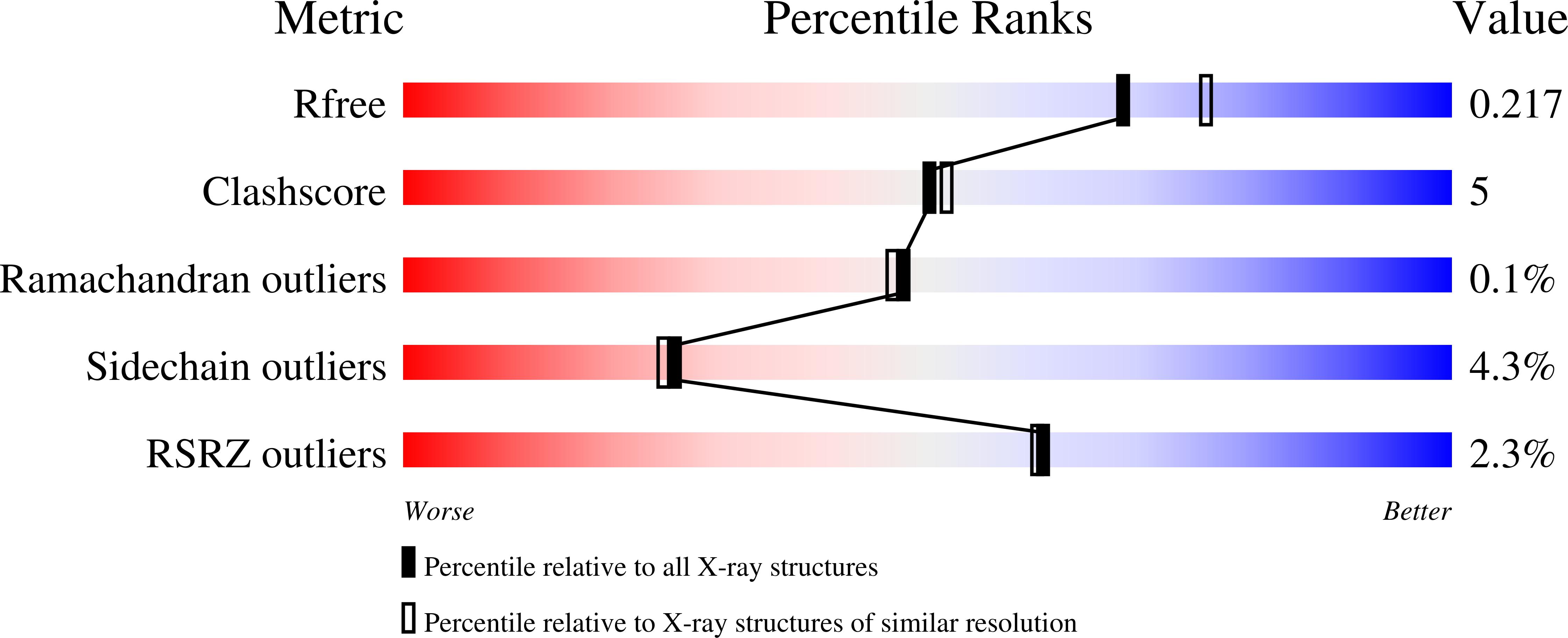
wwPDB Validation report is not available for this entry.
X-ray source:
EMBL/DESY, HAMBURG BEAMLINE BW7B
Spacegroup:
C2
Expression system: Escherichia coli
