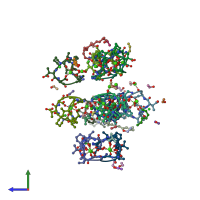
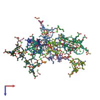
1w3m

X-ray diffraction
1Å resolution

Crystal structure of tsushimycin

Released:
Source organism: Actinoplanes friuliensis
Primary publication:
Structure of the lipopeptide antibiotic tsushimycin.
Acta Crystallogr D Biol Crystallogr 61 1160-4 (2005)
PMID: 16041082

Function and Biology Details

Biochemical function:
  • not assigned
Biological process:
  • not assigned
Cellular component:
  • not assigned

Structure analysis Details

Assembly composition:
homo dimer (preferred)
Assembly name:
PDBe Complex ID:
PDB-CPX-162525 (preferred)
Entry contents:
1 distinct polypeptide molecule
Macromolecule:
TSUSHIMYCIN Chains: A, B, C, D, E, F, G, H, I, J, K, L
Molecule details ›
Chains: A, B, C, D, E, F, G, H, I, J, K, L
Length: 11 amino acids
Theoretical weight: 1.11 KDa
Source organism: Actinoplanes friuliensis

Ligands and Environments

Experiments and Validation Details

Entry percentile scores
X-ray source: BESSY BEAMLINE 14.1
Spacegroup: P1
Unit cell:
a: 33.488Å b: 36.386Å c: 37.511Å
α: 65.64° β: 68.35° γ: 69.88°
R-values:
R R work R free
0.138 not available 0.171