


Function and Biology Details
Biochemical function:
- not assigned
Biological process:
Cellular component:
Structure analysis Details
Assemblies composition:
Assembly name:
Spike protein S2' (preferred)
PDBe Complex ID:
PDB-CPX-145717 (preferred)
Entry contents:
1 distinct polypeptide molecule
Macromolecule:
Spike protein S2'
Molecule details ›
Chains: A, B
Length: 94 amino acids
Theoretical weight: 9.89 KDa
Source organism: Murine hepatitis virus strain A59
Expression system: Escherichia coli
UniProt:
Structure domains: Single alpha-helices involved in coiled-coils or other helix-helix interfaces
Length: 94 amino acids
Theoretical weight: 9.89 KDa
Source organism: Murine hepatitis virus strain A59
Expression system: Escherichia coli
UniProt:
- Canonical:
 P11224 (Residues: 969-1017, 1032-1055, 1056-1059; Coverage: 6%)
Structure domains: Single alpha-helices involved in coiled-coils or other helix-helix interfaces
Ligands and Environments
No bound ligands
No modified residues
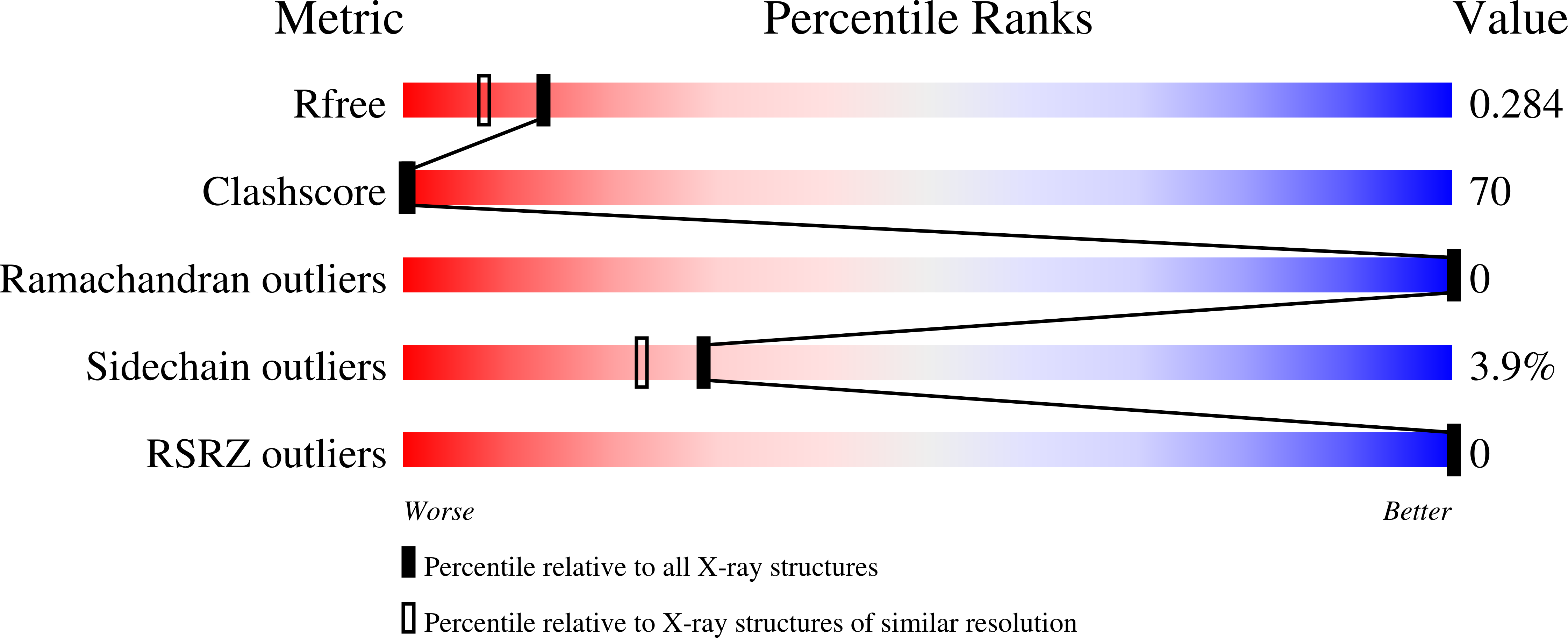
Experiments and Validation Details
wwPDB Validation report is not available for this entry.
X-ray source:
RIGAKU
Spacegroup:
R3
Expression system: Escherichia coli
