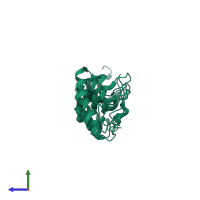
1wg0

X-ray diffraction
2.53Å resolution

Structural comparison of Nas6p protein structures in two different crystal forms

Released:
Source organism: Saccharomyces cerevisiae
Entry authors: Nakamura Y, Umehara T, Tanaka A, Horikoshi M, Yokoyama S, Padmanabhan B, RIKEN Structural Genomics/Proteomics Initiative (RSGI)

Function and Biology Details

Cellular component:

Structure analysis Details

Assembly composition:
monomeric (preferred)
PDBe Complex ID:
PDB-CPX-156123 (preferred)
Entry contents:
1 distinct polypeptide molecule
Macromolecule:
Probable 26S proteasome regulatory subunit p28 Chain: A
Molecule details ›
Chain: A
Length: 243 amino acids
Theoretical weight: 27.39 KDa
Source organism: Saccharomyces cerevisiae
Expression system: Escherichia coli
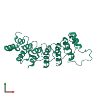
UniProt:
  • Canonical: P50086 (Residues: 1-228; Coverage: 100%)
Gene names: G8564, NAS6, YGR232W
Sequence domains: Ankyrin repeat
Structure domains: Ankyrin repeat-containing domain

Ligands and Environments

No bound ligands
No modified residues

Experiments and Validation Details

Entry percentile scores
X-ray source: SPRING-8 BEAMLINE BL44B2
Spacegroup: P21
Unit cell:
a: 43.444Å b: 68.629Å c: 46.985Å
α: 90° β: 108.95° γ: 90°
R-values:
R R work R free
0.182 0.179 0.25
Expression system: Escherichia coli