


Function and Biology Details
Biochemical function:
Biological process:
- not assigned
Cellular component:
Sequence domains:
Structure domain:
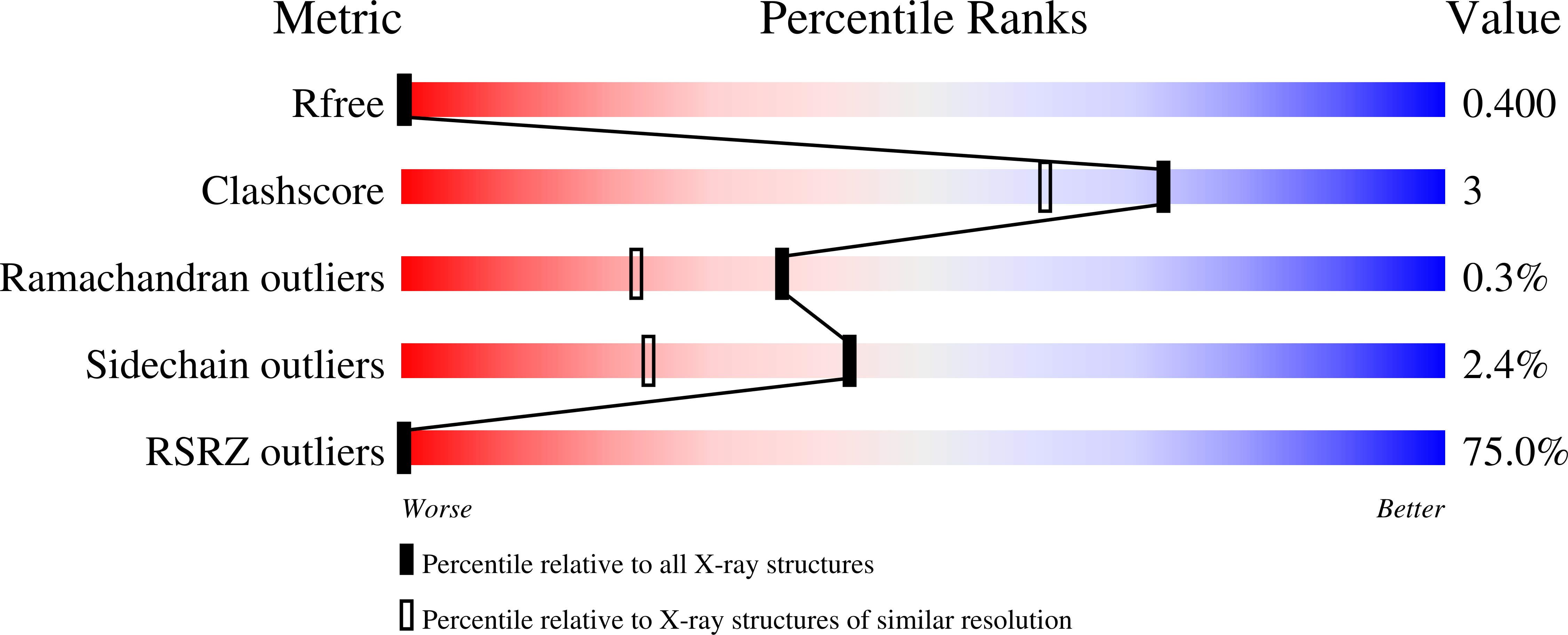
Structure analysis Details
Assemblies composition:
Assembly name:
ABM domain-containing protein (preferred)
PDBe Complex ID:
PDB-CPX-191134 (preferred)
Entry contents:
1 distinct polypeptide molecule
Macromolecule:
ABM domain-containing protein
Molecule details ›
Chains: A, B, C
Length: 99 amino acids
Theoretical weight: 11.47 KDa
Source organism: Pseudomonas aeruginosa
Expression system: Escherichia coli
UniProt:
Sequence domains: Antibiotic biosynthesis monooxygenase
Structure domains: Alpha-Beta Plaits
Length: 99 amino acids
Theoretical weight: 11.47 KDa
Source organism: Pseudomonas aeruginosa
Expression system: Escherichia coli
UniProt:
- Canonical:
 Q9HY51 (Residues: 1-97; Coverage: 100%)
Sequence domains: Antibiotic biosynthesis monooxygenase
Structure domains: Alpha-Beta Plaits


