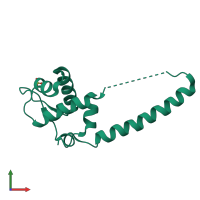
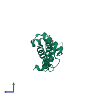
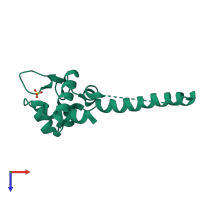
1xd7

X-ray diffraction
2.3Å resolution

Crystal structure of a putative DNA binding protein

Released:
Entry authors: Agarwal R, Swaminathan S, Burley SK, New York SGX Research Center for Structural Genomics (NYSGXRC)

Function and Biology Details

Structure analysis Details

Assembly composition:
homo dimer (preferred)
PDBe Complex ID:
PDB-CPX-159770 (preferred)
Entry contents:
1 distinct polypeptide molecule
Macromolecule:
Putative HTH-type transcriptional regulator YwnA Chain: A
Molecule details ›
Chain: A
Length: 145 amino acids
Theoretical weight: 16.09 KDa
Source organism: Bacillus subtilis subsp. subtilis str. 168
Expression system: Escherichia coli
UniProt:
  • Canonical: P71036 (Residues: 2-133; Coverage: 99%)
Gene names: BSU36630, ywnA
Sequence domains: Iron-dependent Transcriptional regulator
Structure domains: Winged helix-like DNA-binding domain superfamily/Winged helix DNA-binding domain

Ligands and Environments

1 bound ligand:
No modified residues

Experiments and Validation Details

Entry percentile scores
X-ray source: NSLS BEAMLINE X29A
Spacegroup: C2
Unit cell:
a: 65.892Å b: 49.362Å c: 51.51Å
α: 90° β: 121.61° γ: 90°
R-values:
R R work R free
0.26 0.26 0.285
Expression system: Escherichia coli