


Function and Biology Details
Reaction catalysed:
Hydrolysis of the bonds linking certain hydrophobic residues in hemoglobin or globin. Also cleaves small molecules substrates such as Ala-Leu-Glu-Arg-Thr-Phe-|-Phe(NO(2))-Ser-Phe-Pro-Thr.
Biochemical function:
Biological process:
Cellular component:
- not assigned
Sequence domains:
Structure domain:
Structure analysis Details
Assembly composition:
hetero tetramer (preferred)
Assembly name:
Plasmepsin II and peptide (preferred)
PDBe Complex ID:
PDB-CPX-155543 (preferred)
Entry contents:
2 distinct polypeptide molecules
Macromolecules (2 distinct):
Plasmepsin II
Molecule details ›
Chains: A, B
Length: 331 amino acids
Theoretical weight: 37.12 KDa
Source organism: Plasmodium falciparum
UniProt:
Sequence domains: Eukaryotic aspartyl protease
Structure domains: Acid Proteases
Length: 331 amino acids
Theoretical weight: 37.12 KDa
Source organism: Plasmodium falciparum
UniProt:
- Canonical:
 P46925 (Residues: 123-453; Coverage: 73%)
Sequence domains: Eukaryotic aspartyl protease
Structure domains: Acid Proteases
Ligands and Environments
No bound ligands
No modified residues
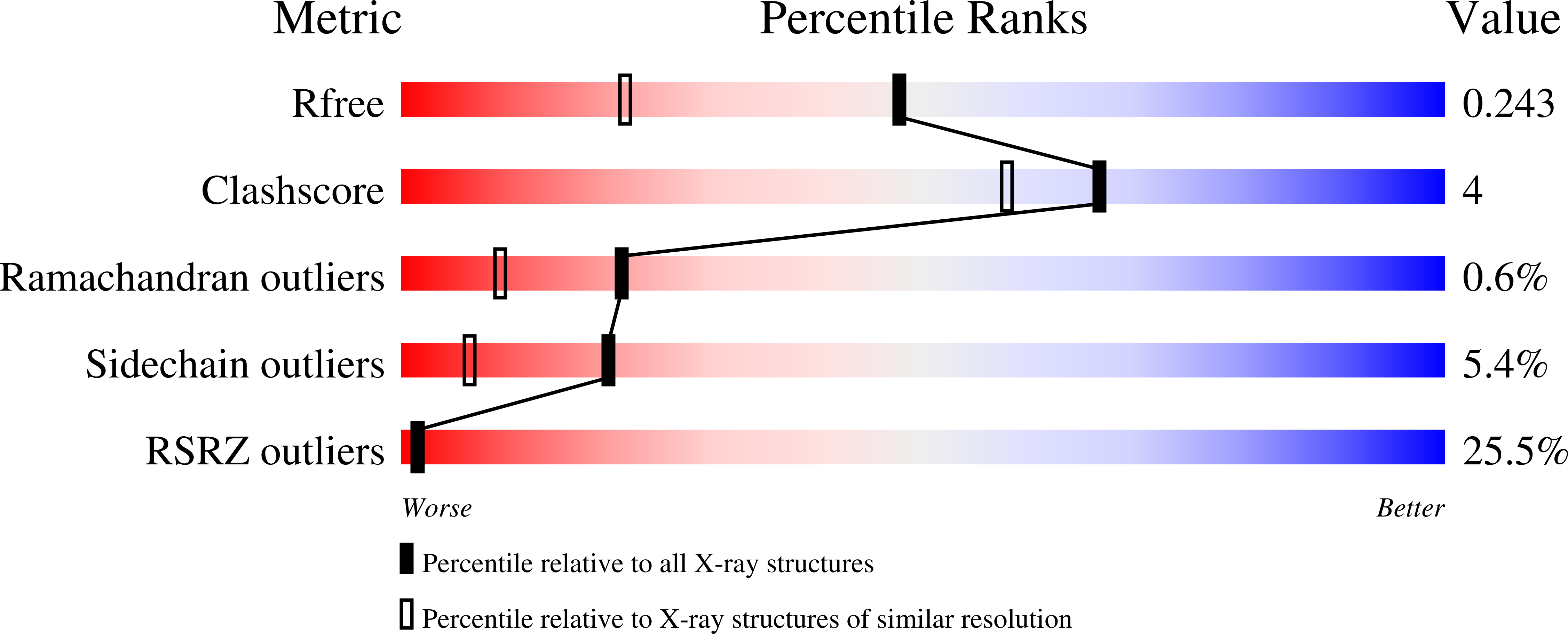
Experiments and Validation Details
wwPDB Validation report is not available for this entry.
X-ray source:
SLS BEAMLINE X06SA
Spacegroup:
P3121
Expression system: Not provided
