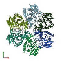
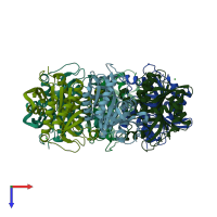
1xe3

X-ray diffraction
2.24Å resolution

Crystal Structure of purine nucleoside phosphorylase DeoD from Bacillus anthracis

Released:
Source organism: Bacillus anthracis
Primary publication:
Structure of purine nucleoside phosphorylase (DeoD) from Bacillus anthracis.
Acta Crystallogr Sect F Struct Biol Cryst Commun 61 459-62 (2005)
PMID: 16511068

Function and Biology Details

Reaction catalysed:
Purine deoxynucleoside + phosphate = purine + 2'-deoxy-alpha-D-ribose 1-phosphate
Biochemical function:
Cellular component:

Structure analysis Details

Assembly composition:
homo hexamer (preferred)
PDBe Complex ID:
PDB-CPX-182596 (preferred)
Entry contents:
1 distinct polypeptide molecule
Macromolecule:
Purine nucleoside phosphorylase DeoD-type Chains: A, B, C, D, E, F
Molecule details ›
Chains: A, B, C, D, E, F
Length: 245 amino acids
Theoretical weight: 26.89 KDa
Source organism: Bacillus anthracis
Expression system: Escherichia coli
UniProt:
  • Canonical: Q81T09 (Residues: 1-235; Coverage: 100%)
Gene names: BAS1372, BA_1483, GBAA_1483, deoD
Sequence domains: Phosphorylase superfamily
Structure domains: Nucleoside phosphorylase domain

Ligands and Environments

1 bound ligand:
No modified residues

Experiments and Validation Details

Entry percentile scores
X-ray source: RIGAKU
Spacegroup: P212121
Unit cell:
a: 63.861Å b: 128.257Å c: 223.565Å
α: 90° β: 90° γ: 90°
R-values:
R R work R free
0.184 0.181 0.235
Expression system: Escherichia coli