


Function and Biology Details
Biochemical function:
Biological process:
Cellular component:
- not assigned
Sequence domains:
Structure domains:
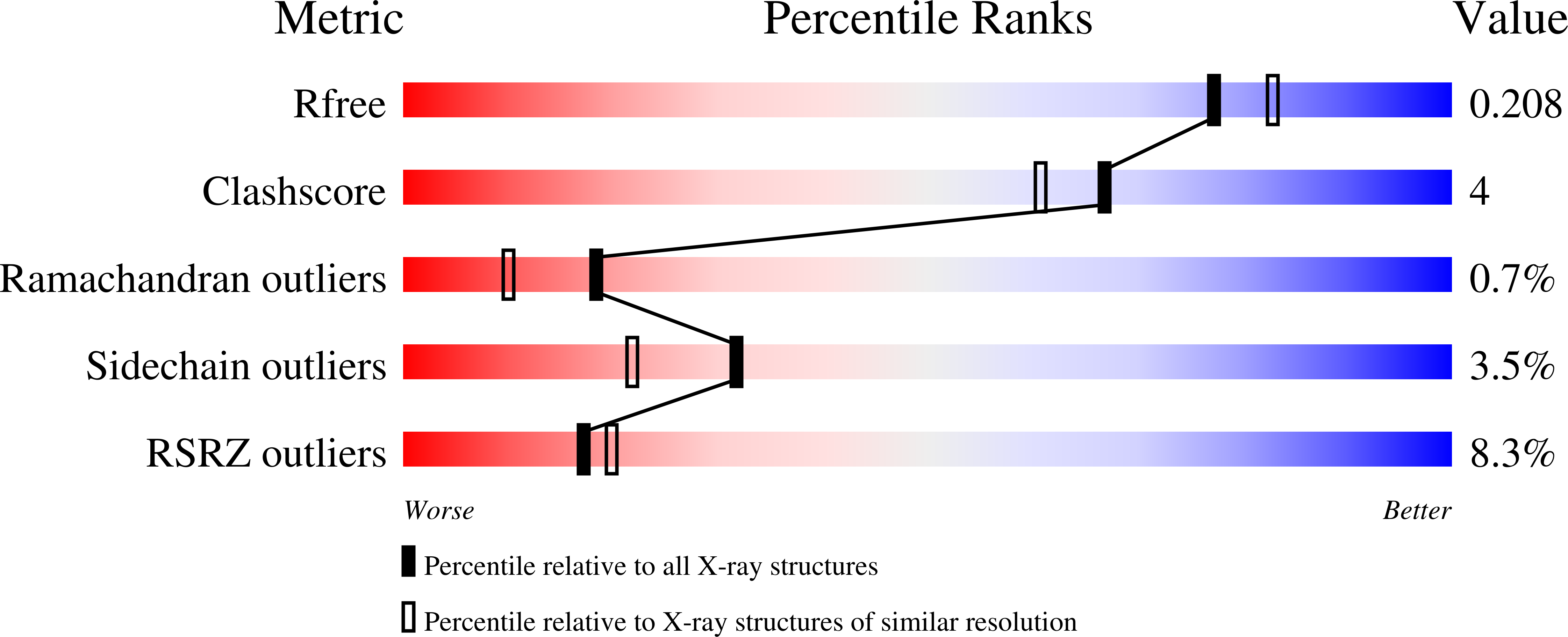
Structure analysis Details
Assembly composition:
homo dodecamer (preferred)
Assembly name:
Tetrahedral aminopeptidase (preferred)
PDBe Complex ID:
PDB-CPX-129829 (preferred)
Entry contents:
1 distinct polypeptide molecule
Macromolecule:
Tetrahedral aminopeptidase
Molecule details ›
Chains: A, B, C, D
Length: 357 amino acids
Theoretical weight: 39.51 KDa
Source organism: Pyrococcus horikoshii
Expression system: Escherichia coli
UniProt:
Sequence domains: M42 glutamyl aminopeptidase
Structure domains:
Length: 357 amino acids
Theoretical weight: 39.51 KDa
Source organism: Pyrococcus horikoshii
Expression system: Escherichia coli
UniProt:
- Canonical:
 O59196 (Residues: 1-353; Coverage: 100%)
Sequence domains: M42 glutamyl aminopeptidase
Structure domains:

