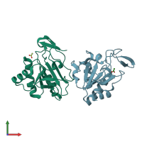
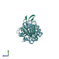
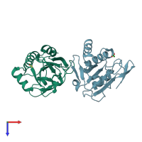
1y25

X-ray diffraction
2.1Å resolution

structure of mycobacterial thiol peroxidase Tpx

Released:
Source organism: Mycobacterium tuberculosis
Primary publication:
Structure of the inactive variant C60S of Mycobacterium tuberculosis thiol peroxidase.
Acta Crystallogr D Biol Crystallogr 62 563-7 (2006)
PMID: 16627951

Function and Biology Details

Reaction catalysed:
Thioredoxin + ROOH = thioredoxin disulfide + H(2)O + ROH

Structure analysis Details

Assembly composition:
homo dimer (preferred)
Assembly name:
PDBe Complex ID:
PDB-CPX-161452 (preferred)
Entry contents:
1 distinct polypeptide molecule
Macromolecule:
Thiol peroxidase Chains: A, B
Molecule details ›
Chains: A, B
Length: 165 amino acids
Theoretical weight: 16.91 KDa
Source organism: Mycobacterium tuberculosis
Expression system: Escherichia coli
UniProt:
  • Canonical: P9WG35 (Residues: 3-165; Coverage: 99%)
Gene names: MTCY09F9.32c, Rv1932, tpx
Sequence domains: Redoxin
Structure domains: Glutaredoxin

Ligands and Environments

1 bound ligand:
No modified residues

Experiments and Validation Details

Entry percentile scores
X-ray source: RIGAKU
Spacegroup: P3121
Unit cell:
a: 106.089Å b: 106.089Å c: 65.334Å
α: 90° β: 90° γ: 120°
R-values:
R R work R free
0.175 0.171 0.249
Expression system: Escherichia coli