


Function and Biology Details
Biochemical function:
Biological process:
Cellular component:
Sequence domains:
Structure analysis Details
Assembly composition:
homo dimer (preferred)
Assembly name:
Disintegrin (preferred)
PDBe Complex ID:
PDB-CPX-177016 (preferred)
Entry contents:
1 distinct polypeptide molecule
Macromolecule:
Disintegrin
Molecule details ›
Chain: A
Length: 64 amino acids
Theoretical weight: 7.14 KDa
Source organism: Echis carinatus
UniProt:
Structure domains: Disintegrin domain
Length: 64 amino acids
Theoretical weight: 7.14 KDa
Source organism: Echis carinatus
UniProt:
- Canonical:
 Q5EE07 (Residues: 1-64; Coverage: 100%)
Structure domains: Disintegrin domain
Ligands and Environments
No bound ligands
No modified residues
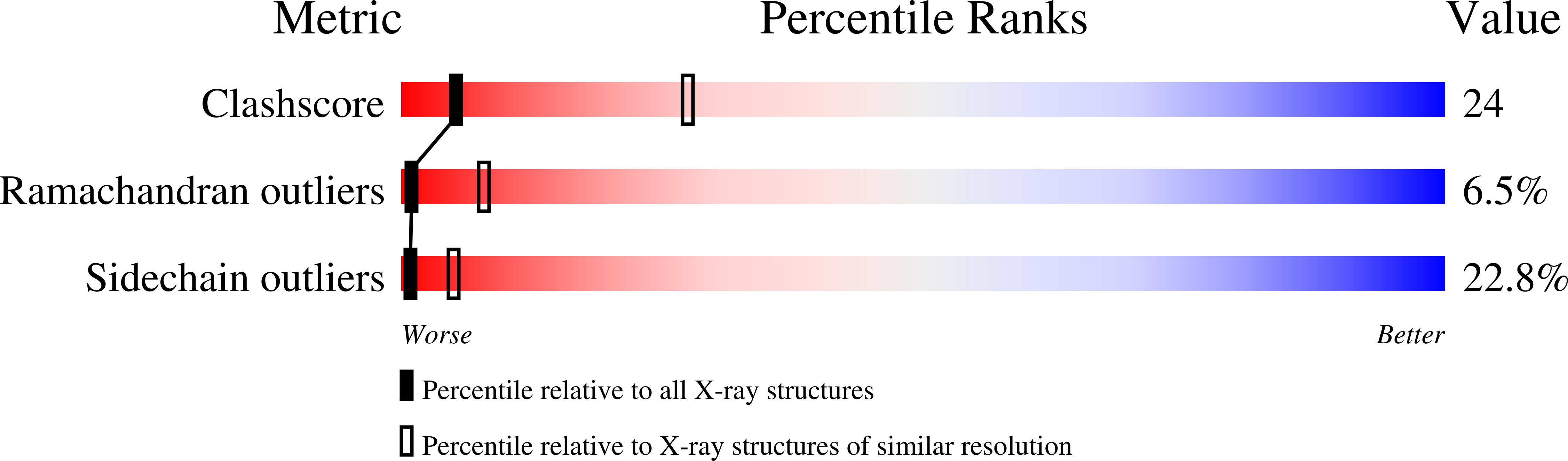
Experiments and Validation Details
wwPDB Validation report is not available for this entry.
X-ray source:
RIGAKU RU300
Spacegroup:
I4122
