1z7k

X-ray diffraction
1.9Å resolution

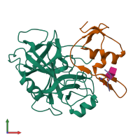
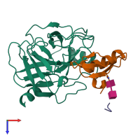
Crystal Structure of Trypsin- Ovomucoid turkey egg white inhibitor complex

Released:
Source organisms:
Primary publication:
Crystal structure of trypsin-turkey egg white inhibitor complex.
Biochem Biophys Res Commun 313 8-16 (2004)
PMID: 14672690

Function and Biology Details

Structure analysis Details

Assembly composition:
hetero trimer (preferred)
Assembly name:
PDBe Complex ID:
PDB-CPX-133387 (preferred)
Entry contents:
3 distinct polypeptide molecules
Macromolecules (4 distinct):
Trypsin Chain: A
Molecule details ›
Chain: A
Length: 223 amino acids
Theoretical weight: 23.49 KDa
Source organism: Sus scrofa
UniProt:
  • Canonical: P00761 (Residues: 9-231; Coverage: 97%)
Sequence domains: Trypsin
Structure domains: Trypsin-like serine proteases
Ovomucoid Chain: B
Molecule details ›
Chain: B
Length: 62 amino acids
Theoretical weight: 6.68 KDa
Source organism: Meleagris gallopavo
UniProt:
  • Canonical: P68390 (Residues: 65-126; Coverage: 34%)
Sequence domains: Kazal-type serine protease inhibitor domain
Structure domains: Wheat Germ Agglutinin (Isolectin 2); domain 1
Ovomucoid Chain: C
Molecule details ›
Chain: C
Length: 4 amino acids
Theoretical weight: 491 Da
Source organism: Meleagris gallopavo
UniProt:
  • Canonical: P68390 (Residues: 12-15; Coverage: 2%)

Ligands and Environments

Carbohydrate polymer : NEW Components: NAG
No bound ligands
No modified residues

Experiments and Validation Details

Entry percentile scores
X-ray source: RIGAKU RU300
Spacegroup: P65
Unit cell:
a: 98.575Å b: 98.575Å c: 73.724Å
α: 90° β: 90° γ: 120°
R-values:
R R work R free
0.172 0.172 0.188