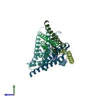
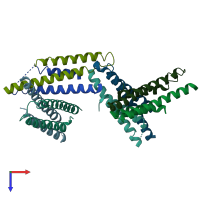
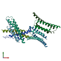
1zw0

X-ray diffraction
1.8Å resolution

Crystal structure of the Yersinia Type III Secretion protein YscE

Released:
Source organism: Yersinia pestis
Primary publication:
Crystal structure of the Yersinia type III secretion protein YscE.
Protein Sci 14 2759-63 (2005)
PMID: 16195558

Function and Biology Details

Biochemical function:
  • not assigned
Biological process:
  • not assigned
Cellular component:

Structure analysis Details

Assemblies composition:
homo dimer (preferred)
homo hexamer
homo tetramer
homo hexadecamer
homo octamer
PDBe Complex ID:
PDB-CPX-130625 (preferred)
Entry contents:
1 distinct polypeptide molecule
Macromolecule:
Type 3 secretion system chaperone YscE Chains: A, B, C, D, E, F, G, H
Molecule details ›
Chains: A, B, C, D, E, F, G, H
Length: 66 amino acids
Theoretical weight: 7.62 KDa
Source organism: Yersinia pestis
Expression system: Escherichia coli
UniProt:
  • Canonical: O68692 (Residues: 1-66; Coverage: 100%)
Gene names: EGX46_00100, Y0027, YPCD1.54, yscE
Sequence domains: Type III secretion system, cytoplasmic E component of needle
Structure domains: Immunoglobulin FC, subunit C

Ligands and Environments

No bound ligands
No modified residues

Experiments and Validation Details

Entry percentile scores
X-ray source: APS BEAMLINE 22-ID
Spacegroup: P21212
Unit cell:
a: 82.969Å b: 84.622Å c: 81.799Å
α: 90° β: 90° γ: 90°
R-values:
R R work R free
0.227 0.225 0.259
Expression system: Escherichia coli