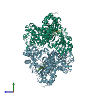
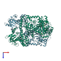
1zxv

X-ray diffraction
2.67Å resolution

X-Ray Crystal Structure of the Anthrax Lethal Factor Bound to a Small Molecule Inhibitor, BI-MFM3, 3-{5-[5-(4-Chloro-phenyl)-furan-2-ylmethylene]-4-oxo-2-thioxo-thiazolidin-3-yl}-propionic acid.

Released:

Function and Biology Details

Reaction catalysed:
Preferred amino acids around the cleavage site can be denoted BBBBxHx-|-H, in which B denotes Arg or Lys, H denotes a hydrophobic amino acid, and x is any amino acid. The only known protein substrates are mitogen-activated protein (MAP) kinase kinases.
Biochemical function:
Biological process:
  • not assigned
Cellular component:

Structure analysis Details

Assembly composition:
monomeric (preferred)
PDBe Complex ID:
PDB-CPX-176027 (preferred)
Entry contents:
1 distinct polypeptide molecule
Macromolecule:
ATLF-like domain-containing protein Chains: A, B
Molecule details ›
Chains: A, B
Length: 776 amino acids
Theoretical weight: 90.36 KDa
Source organism: Bacillus anthracis
Expression system: Bacillus anthracis
UniProt:
  • Canonical: Q52NH3 (Residues: 34-809; Coverage: 96%)
Gene name: lef
Sequence domains:
Structure domains:

Ligands and Environments

2 bound ligands:
No modified residues

Experiments and Validation Details

Entry percentile scores
X-ray source: RIGAKU FR-E
Spacegroup: P21
Unit cell:
a: 95.96Å b: 136.65Å c: 97.9Å
α: 90° β: 98.23° γ: 90°
R-values:
R R work R free
0.231 0.231 0.272
Expression system: Bacillus anthracis