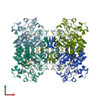
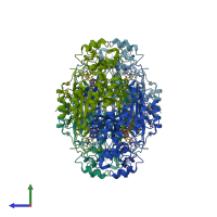
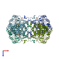
2a5h

X-ray diffraction
2.1Å resolution

2.1 Angstrom X-ray crystal structure of lysine-2,3-aminomutase from Clostridium subterminale SB4, with Michaelis analog (L-alpha-lysine external aldimine form of pyridoxal-5'-phosphate).

Released:
Source organism: Clostridium subterminale
Primary publication:
The x-ray crystal structure of lysine-2,3-aminomutase from Clostridium subterminale.
Proc Natl Acad Sci U S A 102 13819-24 (2005)
PMID: 16166264

Function and Biology Details

Reaction catalysed:
L-lysine = (3S)-3,6-diaminohexanoate
Biochemical function:
Cellular component:
  • not assigned

Structure analysis Details

Assembly composition:
homo tetramer (preferred)
PDBe Complex ID:
PDB-CPX-194986 (preferred)
Entry contents:
1 distinct polypeptide molecule
Macromolecule:
L-lysine 2,3-aminomutase Chains: A, B, C, D
Molecule details ›
Chains: A, B, C, D
Length: 416 amino acids
Theoretical weight: 47.63 KDa
Source organism: Clostridium subterminale
Expression system: Escherichia coli
UniProt:
  • Canonical: Q9XBQ8 (Residues: 1-416; Coverage: 100%)
Gene name: kamA
Sequence domains:
Structure domains:

Ligands and Environments


Cofactor: Ligand PLP 4 x PLP

Cofactor: Ligand SAM 4 x SAM
1 modified residue:

Experiments and Validation Details

Entry percentile scores
X-ray source: NSLS BEAMLINE X25, APS BEAMLINE 17-ID
Spacegroup: C2
Unit cell:
a: 118.89Å b: 92.926Å c: 177.735Å
α: 90° β: 96.74° γ: 90°
R-values:
R R work R free
0.187 0.184 0.225
Expression system: Escherichia coli